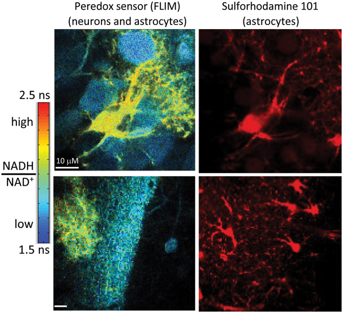
Fig. 13.
Metabolic imaging of the Peredox biosensor of redox, expressed in neurons and astrocytes of acute hippocampal slice. Left: pseudocolor images of the biosensor, which reports high ratio as increased fluorescence lifetime. Right: counterstain with sulforhodamine 101 marks astrocytes specifically and shows that they are the cells with elevated levels in the left-hand images, while neurons have lower levels. Not all cells in the slice express the genetically encoded biosensor, which was introduced with a viral vector. See further discussion of astrocytes in Sec. 3.7.

