Fig. 14.
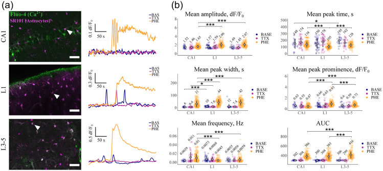
Astrocyte subpopulations in the mouse CNS show significant differences in signaling. transients in sulforhodamine101 (SR101)-labeled astrocytes were detected using Fluo-4. Measurements were made in mouse acute tissue slices containing cortical layer 1 (L1), cortical layers 3-5 (L3-5) and the hippocampal CA1 region (CA1). transients (expressed as fluorescence changes relative to baseline: ) were initially recorded under conditions of baseline activity (BASE) and after sequential application of tetrodotoxin (TTX) (to isolate astrocytes from neuronal activity) and tetrodotoxin plus the -adrenergic receptor agonist phenylephrine (PHE). (a) Representative astrocytes (arrowheads) from three brain areas and the transients recorded from these cells under the various experimental conditions. Scale bar, . (b) Analysis of various transient parameters, recorded under identical experimental conditions. Numerical values are the calculated means for each condition. AUC: area under curve. Modified from Batiuk et al.254 *p ≤ 0.05, **p ≤ 0.01, ***p ≤ 0.001.

