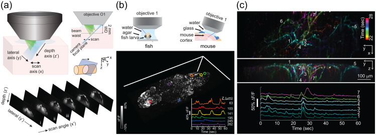
Fig. 20.
SCAPE microscopy: elements and application. (a) Top: SCAPE microscopy uses an oblique light sheet to illuminate the sample, and emitted light is collected by the same objective lens. SCAPE sweeps the oblique light sheet back and forth across the sample, de-scanning and rotating returning light to focus onto a stationary camera. Bottom: example images of a drosophila larva, captured as the light sheet scans. (b) Top: experimental imaging configurations to image neuronal activity in zebrafish larvae and awake, behaving mouse. Bottom: visualization of spontaneous neuronal GCaMP activity in the whole brain of a larval zebrafish. Volume rendering shows a maximum intensity projection over time with inset showing time-courses of 6 cells at 6 different depths in the brain. (c) Top and middle: spontaneous activity of apical dendrites of layer 5 neurons in whisker barrel cortex via GCaMP6f imaging in awake behaving mouse. Maximum intensity projection from the top (XY) and side (YZ) with colors denoting time of peak activity. Bottom: time-courses for the seven numbered regions of interest indicated above.

