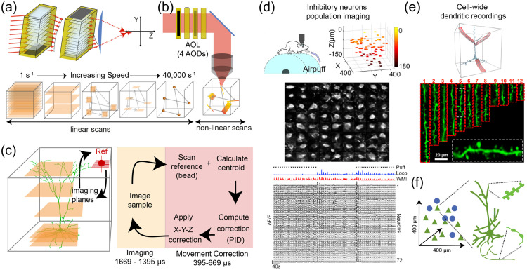
Fig. 24.
Principles and applications of 3D-AOL microscopy. (a) Schematic of pair of AO deflectors (AODs) with chirped sound waves that curve the optical wavefront of a laser beam (red lines). Modified from Ref. 463. (b) Two pairs of orthogonally aligned AODs that make up a spherical AOL that can focus and scan a laser beam in 3D. Cubes below illustrates multiple linear (left) and nonlinear (right) drive-based imaging modes. Modified from Ref. 414. (c) Left: illustration of 3D movement correction with XY and Z scans of a bead interleaved with multiplane imaging. Right: sequence of operation to track a reference object in 3D and compensate imaging for brain movement. Modified from Ref. 462. (d) Selective imaging of sparsely distributed inhibitory interneurons in the cerebellum. Top left: awake mouse on a treadmill. Top right: locations of somatic ROIs in 3D-AOL imaging volume of mouse expressing GCaMP6f in Golgi cells. Middle: montage of 72 selectively imaged Golgi cell somata. Bottom: activity traces extracted from the cells during locomotion, whisking (WMI) and mild air puff to the whiskers (Puff). Modified from Ref. 465. (e) Image of pyramidal cell with dendritic branches selectively imaged with multiple line scans (Ribbon scanning). Aligned images of individual dendritic segments with spines clearly visible (middle). Modified from Ref. 466. (f) Schematic illustrating multiscale imaging functionality of 3D-AOL microscopy, which enables simultaneous imaging of neuronal populations, dendrites, spines and axons present in a local circuit.

