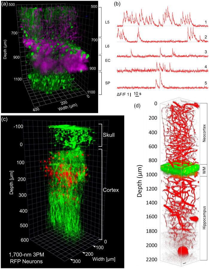
Fig. 27.
Deep imaging with 3PM. (a) 3PM of GCaMP6s-labeled neurons in the mouse cortex and the hippocampus. Green, fluorescence; magenta, third-harmonic generation (THG). (b) Activity recording in the Stratum Pyramidale layer of the hippocampus at depth. Only a small portion of the 48-minute recording session is shown. Reproduced with permission from Ref. 416. Springer Nature. (c) Imaging through the intact mouse skull. Red, RFP-labeled neurons in a Brainbow mouse, male, 12 weeks. Green, THG. The zero depth is set just beneath the skull. Reproduced with permission from Ref. 495. Springer Nature. (d) In vivo 3PM of mouse brain vasculature labeled with quantum dots (Qtracker 655) to depth. Reproduced with permission from Ref. 496, © 2019 American Chemical Society.

