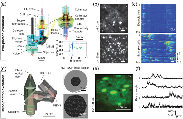
Fig. 28.
2P and 3P wearable microscopes for cellular-resolution imaging in freely behaving animals. (a) Example 2P miniscope for high-resolution imaging at depth. This implementation weighs 4.2 grams and includes an electrically tunable lens (ETL) (top right) for rapid focus adjustment across a focal range (bottom right). Using a microelectromechanical system (MEMS) scanner, the miniscope allows 10 Hz recordings at 512 x 512 pixels resolution across a FOV. (b) Example images of GCaMP6s-expressing neurons acquired at the indicated focal depths (z) in the mouse prefrontal cortex. (c) transients from dendritic (top) and cell body regions of interest (ROIs) (bottom) within the focal planes shown in (b). (d) 3P miniscope for deep, high-resolution imaging. This microscope weighs 5 grams and includes a 1.2-m hollow-core photonic bandgap crystal fiber (HC-PBGF) (right) for low-dispersion delivery of the excitation light-pulses. A plastic optical fiber collects the emitted fluorescence for remote detection by photomultiplier tubes (PMTs). (e) Image of GCaMP6s-labeled rat cortical neurons at the indicated focal depth (z) in the posterior parietal cortex. (f) spiking from four example neurons recorded at cortical depth. (a)–(f) Adapted with permission from Nature Publishing Group: (a)–(c) from Ref. 418 and (d)–(f) from Ref. 417.

