Fig. 3.
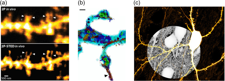
Super-resolution imaging of fine neuronal structures. (a) Comparison of 2P versus 2P-STED image of dendritic spines imaged in the mouse hippocampus in vivo (adapted from Ref. 36). (b) Correlative STED and SMLM image of dendritic spine morphology and dynamic spatial arrangement of synaptic proteins (blue, neuronal morphology; green, PSD-95 scaffold protein; colored tracks, GluA1 receptor subunit), scale bar 500 nm (adapted from Ref. 37). (c) Super-resolution shadow imaging (SUSHI) of brain tissue, revealing anatomical context of a YFP-labeled neuron (adapted from Ref. 38).

