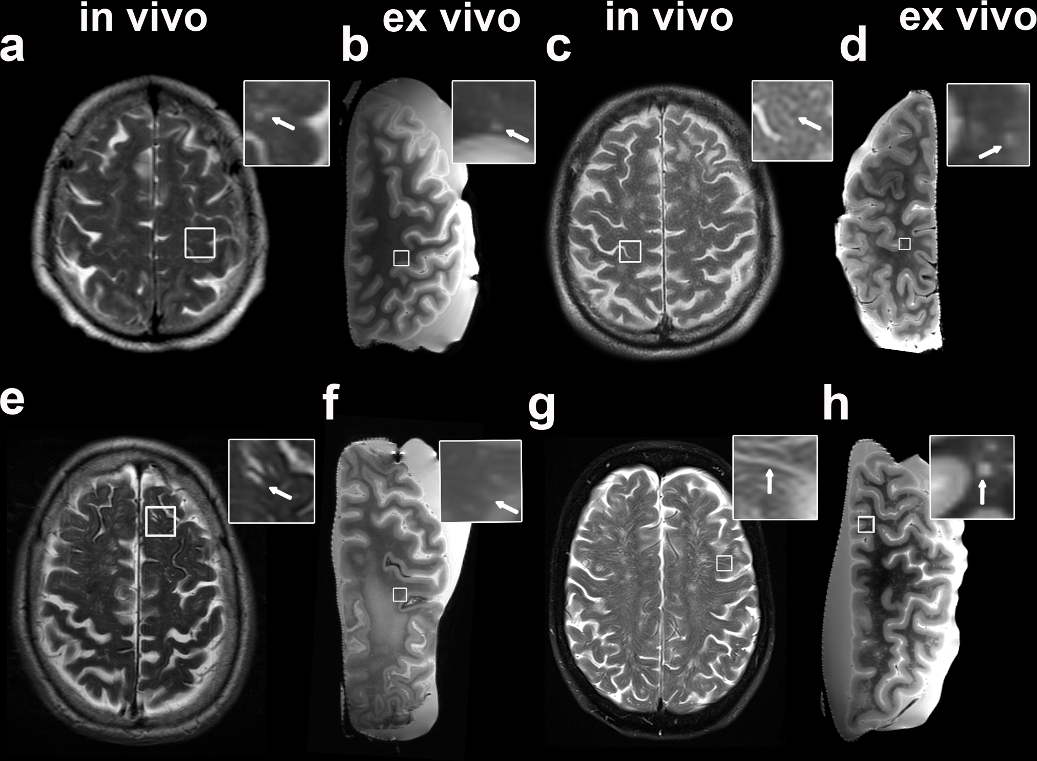
Fig. 1.

Examples of MRI-visible perivascular spaces (PVS) on in vivo and ex vivo MRI. The figure shows both examples of clinical in vivo (a, c, e, g) and of ex vivo (b, d, f, h) T2-weighted MRI scans of the same patient. The first is a case with mild degree of MRI-visible PVS in vivo and frequent after death (score in vivo 1 (a), ex vivo 3 (b); In vivo MRI-death interval: 7 months); the second case displays moderate MRI-visible PVS (score in vivo 2 (b), ex vivo 2 (c); In vivo MRI-death interval: 8 months). In the bottom row, a case with in vivo frequent and ex vivo severe (score in vivo 3 (e), ex vivo 4 (f); In vivo MRI-death interval: 53 months) and finally one with severe (score in vivo 4 (g), ex vivo 4 (h); In vivo MRI-death interval: 67 months) MRI-visible PVS are displayed. The insets show details of MRI-visible PVS (arrows)
