Fig. 2.

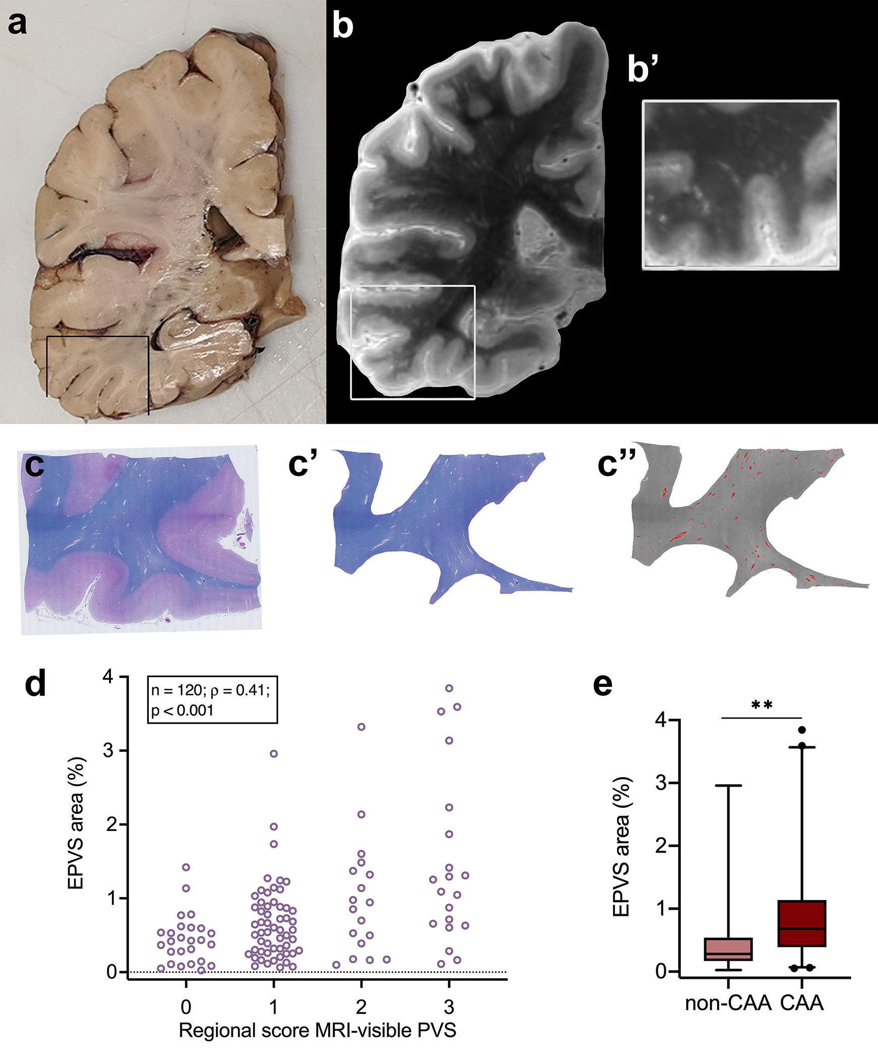
Quantification of enlarged perivascular spaces (EPVS) on ex vivo MRI and histopathology. Example of an approximately 1 cm thick coronally cut slab (a). The box highlights the region sampled for histopathology. Coronal view of the 3 tesla ex vivo turbo spin echo sequence of the same hemisphere at the corresponding level (b). The inset shows a higher magnification of the same area at the level assessed on histopathology for EPVS severity (b’). Luxol fast blue with Hematoxylin&Eosin (LHE)-stained section from the same sample (c), with corresponding manual segmentation of the white matter (c’) and semi-automatic segmentation of the EPVS (overlaid in red) (c”). Positive correlation between the regional score of MRI-visible PVS and the percentage area of EPVS of the total white matter area, calculated on histopathological sections (one from the basal ganglia and four from cortical regions) of cases with and without CAA (d). EPVS percentage area in CAA cases was significantly higher than in non-CAA cases in the four cortical regions (frontal, temporal, parietal, and occipital). ** = p < 0.01 (e)
