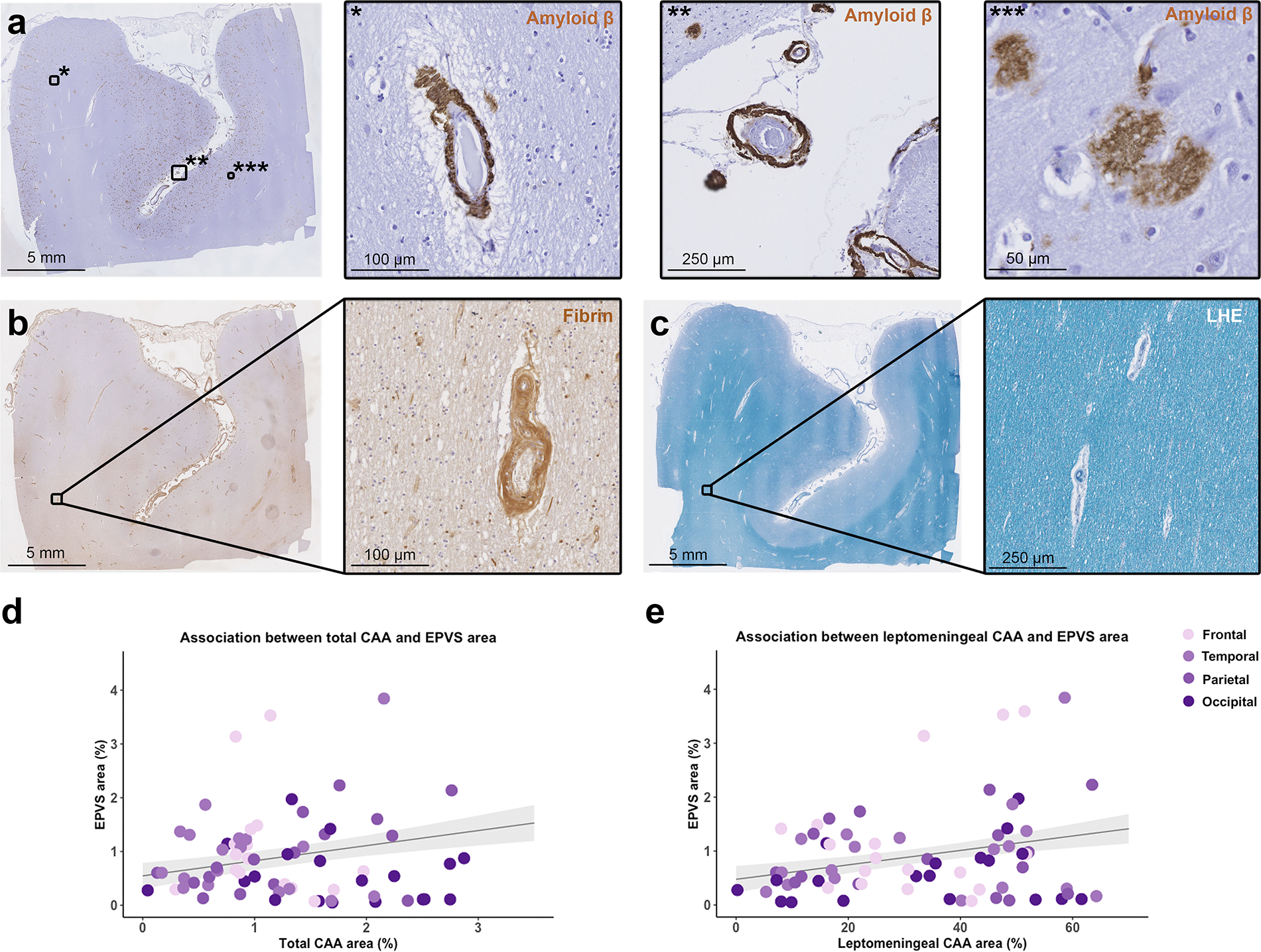
Fig. 3.

Correlation between enlarged perivascular spaces (EPVS) and cerebral amyloid angiopathy (CAA). The figure shows representative examples of the histopathological markers included in the analysis. Low magnification overview of a section that underwent immunohistochemistry against Aβ (a) and details of cortical CAA (*), leptomeningeal CAA (**) and Aβ plaques (***); an adjacent section that underwent immunohistochemistry against fibrin (b) and detail of a fibrin positive vessel in the white matter; a further adjacent section stained for Luxol fast blue with Hematoxylin&Eosin (LHE) (c) with detail of a portion of the white matter. Graphs representing the positive significant association between total CAA percentage area and EPVS percentage area in the cortical regions of CAA cases (d), and between leptomeningeal CAA percentage area and EPVS percentage area (e) (n = 19 cases; n = 72 sections; grey shadowed area shows the standard error of the model’s prediction)
