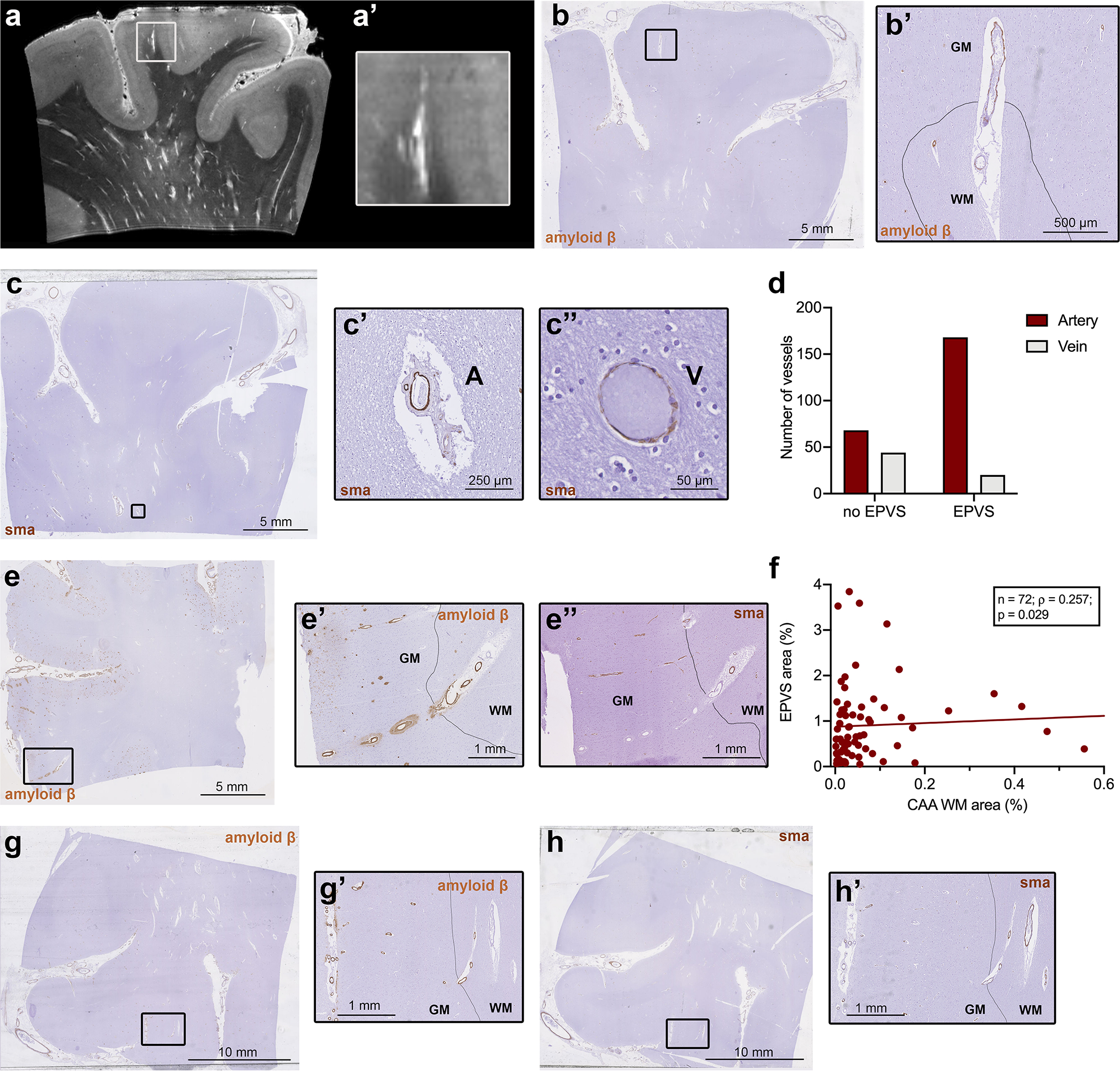
Fig. 5.

Histopathological characterization of individual blood vessels with surrounding enlarged perivascular spaces (EPVS). Ultra-high resolution 7 tesla turbo spin echo scan of case no. 13, with an example of an EPVS extending into the cortex (a, a’). The corresponding histopathological section, on which the drawn black line represents the boundary between grey matter (indicated by GM) and white matter (indicated by WM), confirms this observation (b, b’). On serial sections immunohistochemistry for smooth muscle actin (SMA) was performed (c) and revealed, that the vast majority of vessels with an EPVS were arterioles (χ2(1) = 28.29; n = 280; p < 0.001) (d), suggesting that EPVS are mainly peri-arteriolar (c’, A = artery) and not peri-venular (c”, V = vein). On some Aβ stained cortical sections, Aβ was present not only in the wall of the cortical portion of the vessel, but to a minor extent, also in the white matter portion of the same vessel (examples are b’, e’ and g’). The same vessels showed severe loss of SMA in the cortex, but not in the WM (e”, h’). A significant positive association was found between the percentage area of EPVS and the percentage area occupied by cerebral amyloid angiopathy (CAA) in the white matter, within all CAA cases (n=72 samples in 19 cases) (f)
