
Fig. 6.
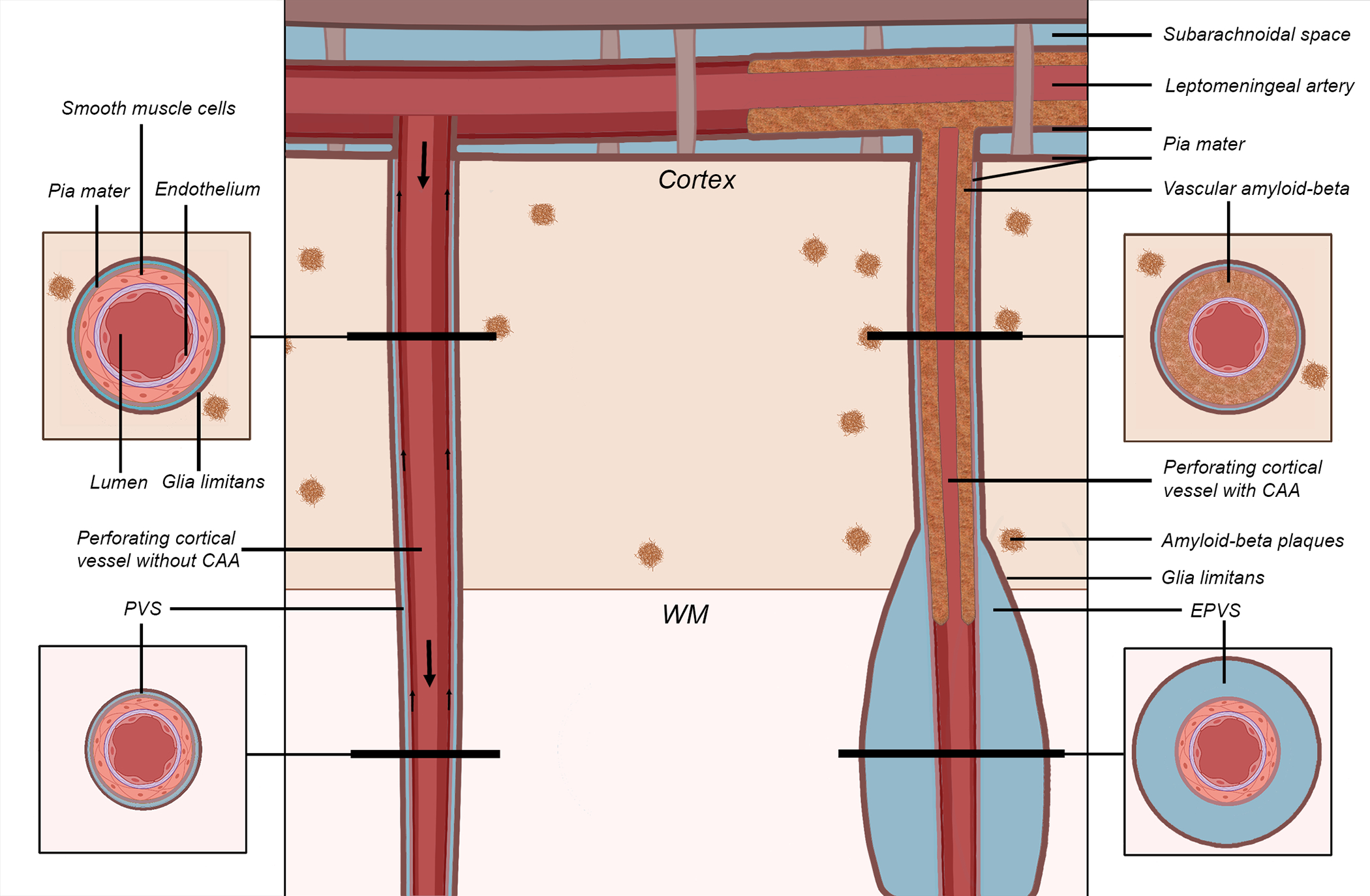
Schematic representation of the proposed mechanism of perivascular space (PVS) enlargement in cerebral amyloid angiopathy (CAA). The vessel on the left represents a healthy perforating artery (in red). The vessel on the right represents a perforating artery affected by CAA, in which Aβ accumulates within the tunica media of the wall of the cortical portion of the vessel (in brown), replacing the smooth muscle cells and leading to an enlargement of the PVS in the white matter portion of the same vessel (giving rise to the hypothesized self-reinforcing mechanism of continuing vascular Aβ accumulation). The smaller arrows on the left indicate the presumed direction of Aβ clearance along the vessel (either along the basement membranes or the perivascular compartment), while the bigger arrows represent the direction of blood flow. The results lend support to this proposed model of perivascular Aβ clearance. Key: CAA = cerebral amyloid angiopathy; EPVS = enlarged perivascular space; WM = white matter