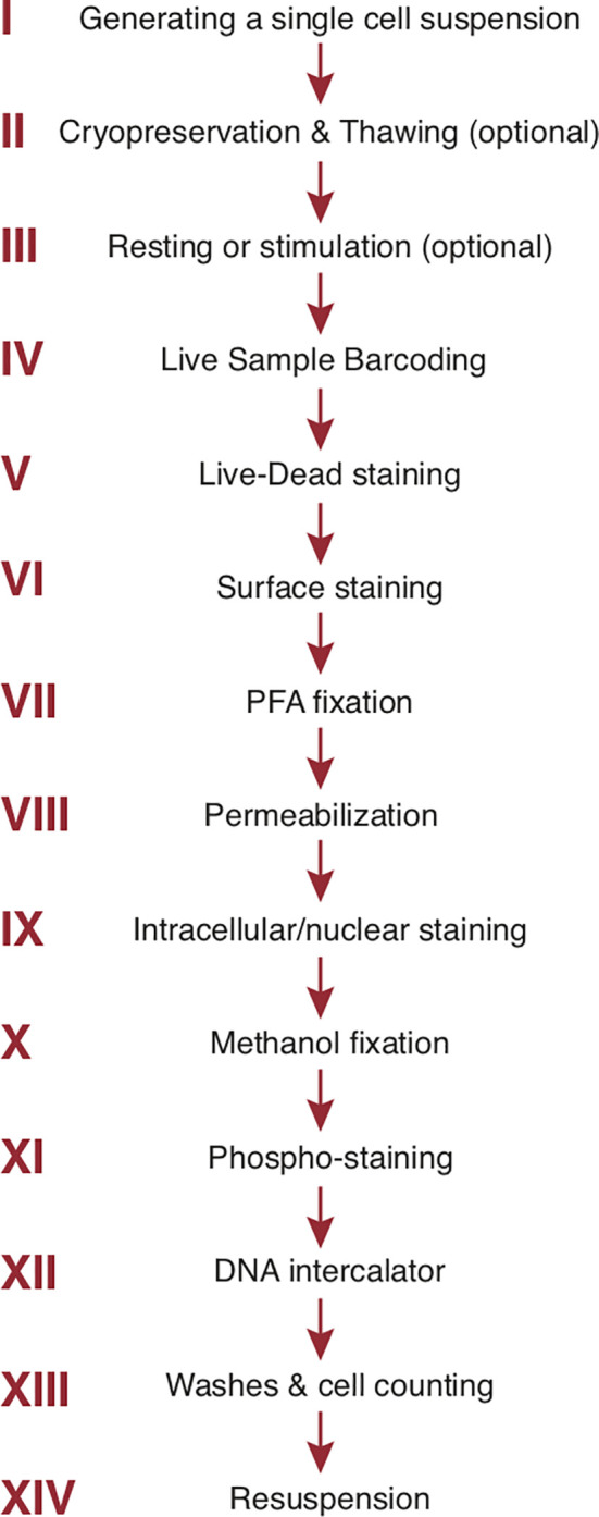
Figure 3.

Step-by-step sample preparation. Representative sample preparation protocol. The order of individual steps may vary depending upon the experimental design (see text). Sample washes with cell staining buffer separate each step. The final 3 steps are specific to mass cytometry. PFA, paraformaldehyde.
