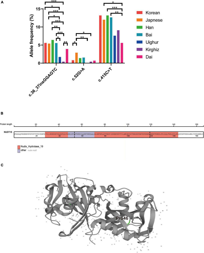
FIGURE 3.
Comparison of the frequencies of variation at multiple sites of NUDT15 in different ethnic groups and the NUDT15 structural domain and protein structure. (A) *P < 0.05; **P < 0.01; ***P < 0.001. (B) The NUDT15 structural domain obtained from St. Jude Cloud is marked in color, and p.P46R (c.137C > G and c.138T > G) was located on the Nudix-Hydrolase-19 functional domain. (C) The position of p.P46R in the NUDT15 protein derived from Uniprot is highlighted in green.

