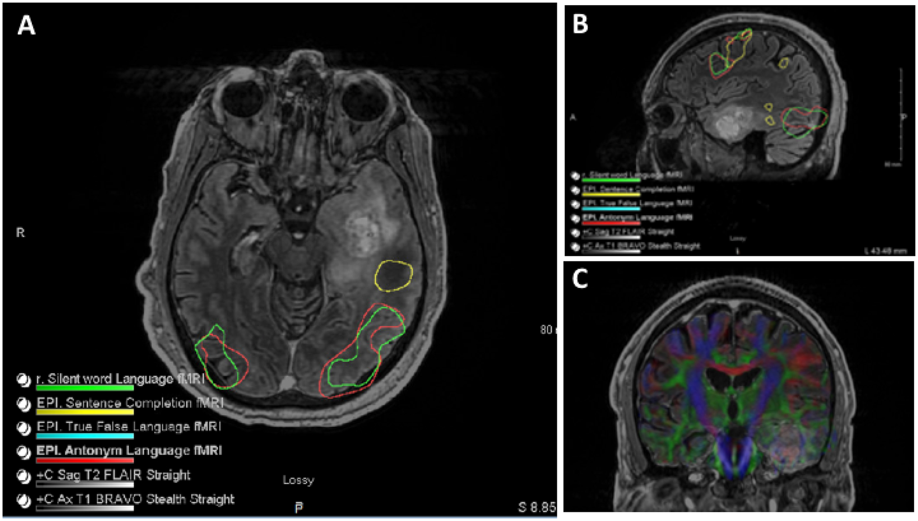
Figure 2: fMRI and DTI imaging:

A) Axial & B) sagittal FLAIR sequencing demonstrating left temporal lesion with functional mapping. C) Tractography shows the relationship of major tracts in relation to the lesion.

A) Axial & B) sagittal FLAIR sequencing demonstrating left temporal lesion with functional mapping. C) Tractography shows the relationship of major tracts in relation to the lesion.