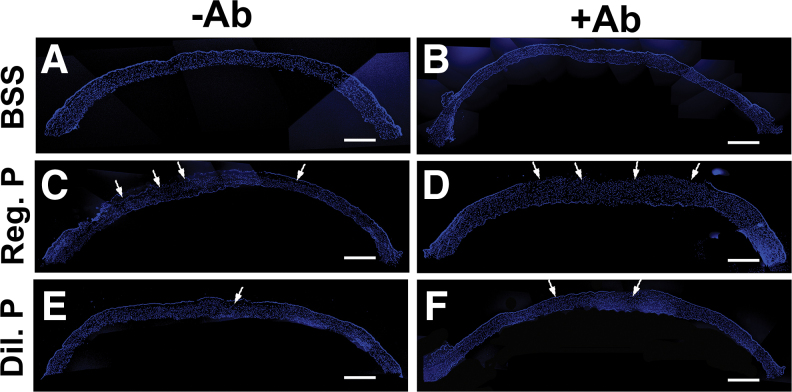
FIG. 6.
Proparacaine effects on corneal reepithelialization and stromal cell density in vivo. The panoramic images of DAPI immunofluorescence staining of whole tissue sections of rabbit cornea show the reepithelialization pattern with BSS without antibiotics (A), BSS with antibiotics (B), regular proparacaine without antibiotics (C), regular proparacaine with antibiotics (D), diluted proparacaine without antibiotics (E), and diluted proparacaine with antibiotics (F) in corneal tissue sections. Regular strength proparacaine delayed the reepithelialization process compared with diluted strength of proparacaine at day 3. Arrows show the cellular density. Scale bar = 100 μm. DAPI, 4′,6-diamidino-2-phenylindole. Color images are available online.

