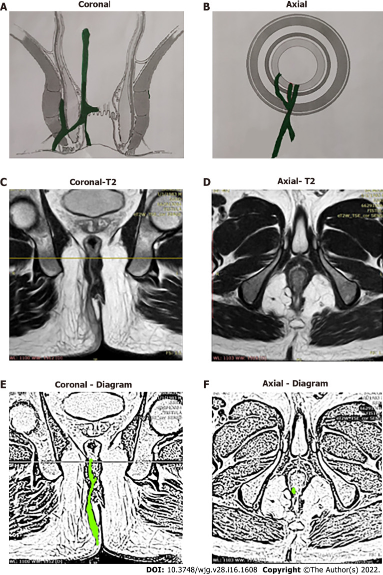
Figure 9.
A 36-year-old male patient with a high intrarectal fistula at 7 o’clock. A: Coronal section (schematic diagram); B: Axial section (schematic diagram); C: T2-weighted magnetic resonance imaging (MRI) Coronal section showing high intrarectal fistula tract; D: T2-weighted MRI Axial section; E: Sketch of MRI Coronal section highlighting high intrarectal fistula tract (light green color); F: Sketch of MRI Axial section highlighting fistula tract (light green color).

