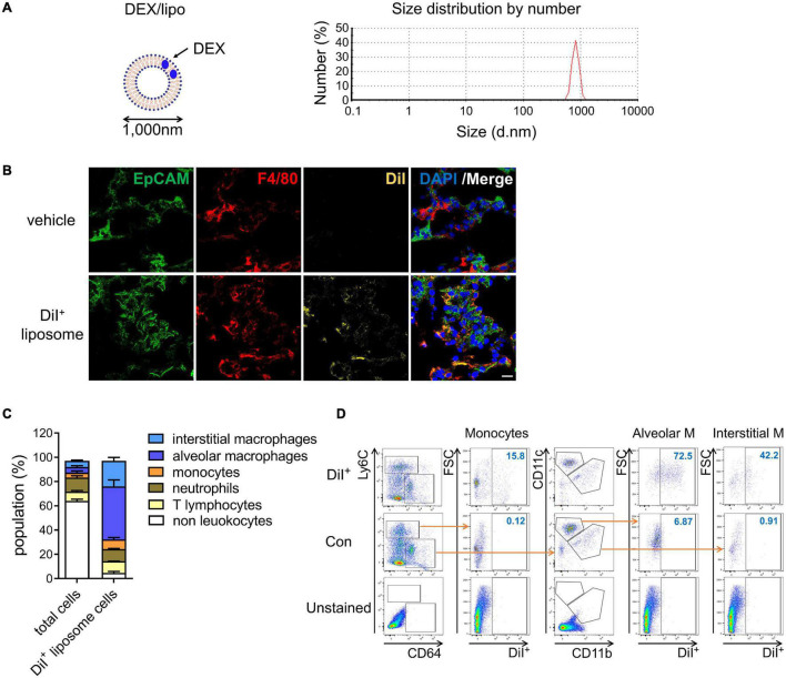
FIGURE 1.
DEX/lipo is specifically delivered into the macrophages in lungs. (A) Schematic of DEX/lipo, left, and size determination of DEX/lipo by dynamic light scattering (DLS), right. The mean diameter is close to 1,000 nm. (B) Co-immunofluorescence (Co-IF) staining was performed to confirm the co-localization of the epithelial cell adhesion molecule (EpCAM) (green) or F4/80 (red) with DiI (yellow). Mice were injected intranally with 20 μl of DiI-labeled liposomes, and lung tissues were dissected at 24 h post-injection. Scale bar; 20 μm. (C) Population percentages in the lungs of mice are shown as total cells compared to DiI+ cells. Representatives of two independent experiments are shown. (D) Flow-cytometry analysis of DiI+ cells. CD45+Ly6C+CD64low monocytes population, CD45+Ly6C–CD64+CD11c+ alveolar macrophages population, and CD45+Ly6C–CD64+CD11b+ interstitial macrophages population are shown in the DiI/forward scatter (FSC) dot plot with gating for the DiI+ population.

