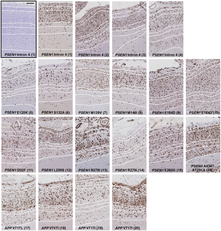
FIGURE 2.

Representative images of Aβ immunohistochemically stained frontal cortex of each individual case within the mutation sub‐groups, PSEN1 pre‐codon 200 (10 cases), PSEN1 post‐codon 200 (6 cases) and APP (4 cases). One Nissl stained PSEN1 Intron 4 mutation tissue section is shown (black box) to highlight how the layers were defined based on cellular morphology and applied to the serial Aβ section. The 6 cortical layers are defined by white lines, with layer 1 at the pial surface and layer 6 adjacent to the white matter. Black scale bar =500µm. Numbers in brackets refer to case number.
