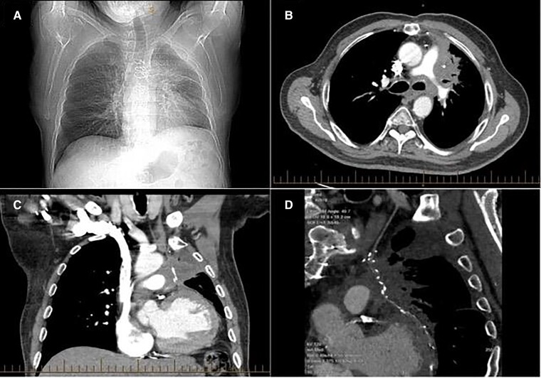
Figure 1.
Chest X-ray and computerized tomography chest at admission demonstrating infiltrating mass in the left lung. The chest X-ray in (A) demonstrates hyperinflation of the lungs with increased peribronchial markings in the left lung. The computerized tomography demonstrates in panel (B), an infiltrative irregular and speculated mass in the left hilum measuring ∼10 cm × 6.5 cm. There is peribronchovascular thickening and compression of the left upper lobe bronchus with complete occlusion of the small airways. There is involvement of the left pleura (B) and (C). The mass is encasing the left internal mammary artery graft, with resultant extrinsic compression (C) and (D).

