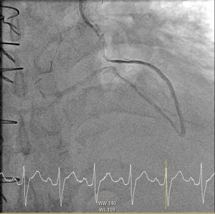
Figure 3.
Diagnostic coronary angiogram demonstrating compression of the left internal mammary artery to obtuse marginal graft. This is a LAO 2.8° and cranial 27.2° view with the left internal mammary artery graft selectively engaged. There is a diffuse segment of flow-limiting disease in the mid-section of the left internal mammary artery to obtuse marginal graft with flow distal to the lesion, secondary to extrinsic compression of the tumour. There is no atherosclerotic disease in the rest of the graft.

