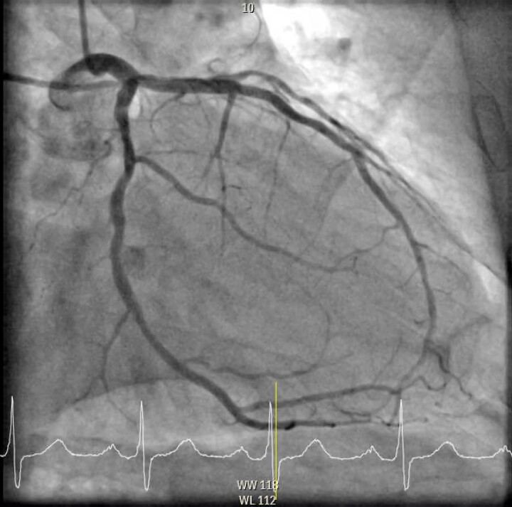
Figure 4.
Final image of left circumflex artery after the intravascular ultrasound-guided percutaneous coronary intervention of the chronic total occlusion. In the LAO 49.7° and caudal −15.0° view with engagement of the left main coronary artery showing flow restored to a revascularized proximal left circumflex artery after the intravascular ultrasound-guided percutaneous coronary intervention with intravascular lithotripsy and placement of two drug-eluting stents (Osiro 3.0 × 22 mm + 2.5 × 26 mm).

