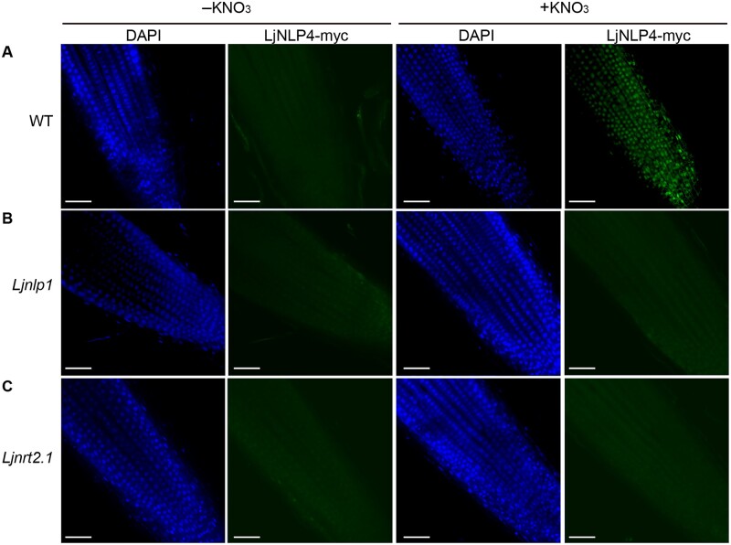
Figure 7.
Subcellular localization of LjNLP4. A–C, Immunohistochemistry of the LjNLP4-myc protein in root apical cells of WT, Ljnlp1, and Ljnrt2.1-1 mutants. A monoclonal anti-myc antibody and an antibody conjugated to Alexa Fluor 488 Plus (right: green signal) were used as the primary and secondary antibodies. Nuclei were visualized with DAPI (left: blue signal). Plants with transgenic hairy roots transformed with the LjUBQpro:LjNLP4-myc construct were grown on a nitrogen-free medium for 3 days and then supplied with 0 (−) or 10 mM KNO3 (+) for 1 h. Representative images of at least five independent locations are shown for each condition. Scale bars = 50 µm.

