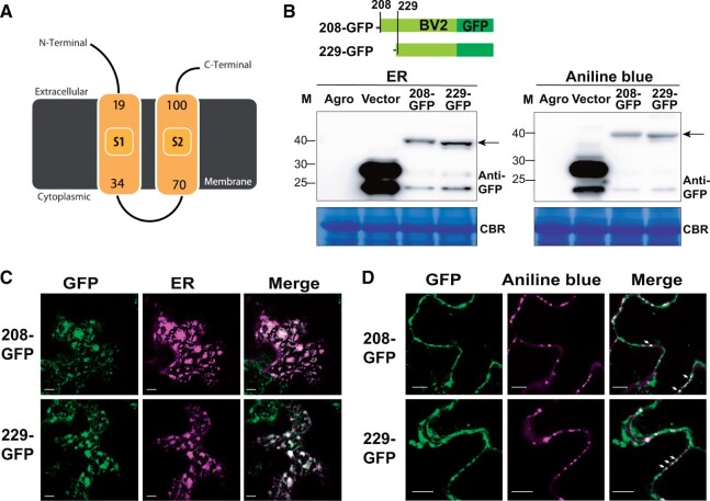
Figure 6.
Subcellular localization of BV2 in the ER and PD. A, Plot of BV2 protein structure showing the predicted transmembrane domains (yellow) analyzed and generated via Phypy2 (http://www.sbg.bio.ic.ac.uk/phyre2). B–D, As described in Figure 3, H and I, but for the protein expression (B) and subcellular localization (C, D) of the GFP-tagged BV2 protein initiated from AUG208 or AUG229. ER marker: CD3-959 (Nelson et al., 2007). Aniline blue: a PD dye. Scale bar = 10 µm.

