. 2020 Jan 23;10(7):3884–3894. doi: 10.1039/c9ra08203d

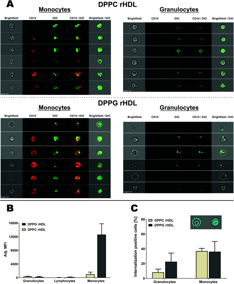
Fig. 8. Analysis of rHDL interaction with leukocytes using imaging flow cytometry for both DPPC and DPPG rHDL. (A) Selected DiO positive cells from two donors shown with brightfield (Ch01), CD14 (Ch11), DiO (Ch02), CD14/DiO and brightfield/DiO. The same contrast settings were used for monocytes and granulocytes for each of the rHDL formulations. (B) The imaging flow cytometry confirmed the preferred monocyte association of rHDL. The MFI was subtracted background from the specific cell type and adjusted by the integral of DiO absorbance from the SEC analysis of each rHDL formulation. (C) The brightfield images were used to define masks that defined the interior or the cells, as illustrated for a single monocyte in the inset, which also shows a mask of the cell surface. This allowed for the determination of the internalization of rHDL cargo, which showed more internalization positive monocytes than granulocytes. The data in B and C represent mean + SEM (n = 2).

Fig. 8