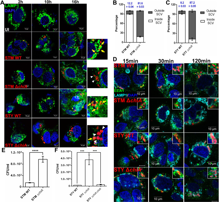
Fig 3. ΔchiA mutants quit SCVs in the epithelial cells and hyper-proliferates in the cytoplasm.
(A) Representative image of Caco-2 cells infected with STM WT, STM ΔchiA, STY WT and STY ΔchiA strains to visualize the intracellular niche of the bacteria. The SCVs were stained for LAMP1 (UI- Uninfected; Yellow arrows- LAMP1+ SCVs, white arrowheads- LAMP1- SCVs). (B) % of STM WT and STM ΔchiA, (C) % of STY WT and STY ΔchiA bacteria inside and outside the LAMP1+ SCVs 16 hpi was calculated. (N = 3). (D) Representative image of Caco-2 cells infected with STM WT, STM ΔchiA, STY WT and STY ΔchiA strains at MOI 50 to visualize EEA1 and LAMP1 recruitment on the SCVs (scale bars- 10μm; insets show SCVs). Absolute CFU/ml values of (E) STM WT and STM ΔchiA, and (F) STY WT, STY ΔchiA and STY ΔchiA:chiA in Caco-2 cells in chloroquine resistance assay 16 hpi. (N = 3, n = 3). One-way ANOVA was used to analyze the data.

