. 2020 Feb 11;10(11):6306–6314. doi: 10.1039/c9ra10385f

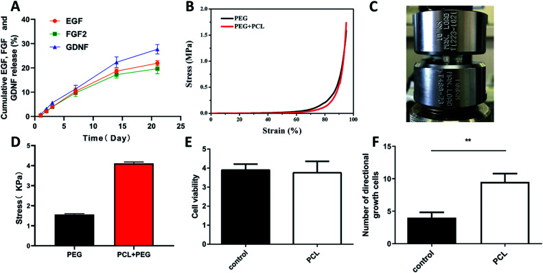
Fig. 3. Slow-release, mechanical, and biocompatibility of cytokine-loaded PCL-PEG scaffold composite. (A) the cumulative release of EGF, FGF2 and GDNF from the PEG hydrogels, as determined by the ELISA kits; (B–D) the cyclic compression stress–strain curves of PEG hydrogels and PCL-PEG scaffold composite and the comparison between them; (E) bar graph showing the proliferation of PC12 cells with PCL co-culture or not by using CCK8 assay on day 3 (n = 6); (F) comparison of the number of directional adhesion cells between the two groups. *P < 0.05, **P < 0.01, One-Way ANOVA followed by Tukey's post hoc test. N. S. indicates no significant difference.

Fig. 3