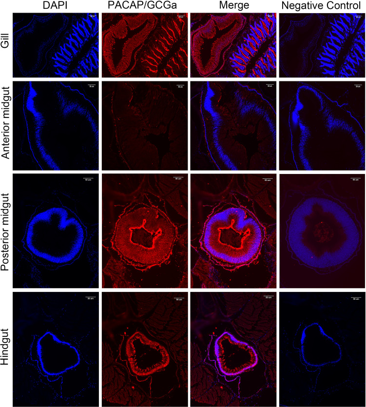
Figure 3.
Detection of PACAP/GCGa in several adult B. belcheri tissues by immunofluorescence. Immunofluorescence digital images of DAPI staining (blue), PACAP/GCGa (red) positive signals, and merged and negative control (merged). Negative controls represent the result of incubations in which primary antibody with overnight incubation with antigen in 1:10 molar ratio was used to replace primary antibody alone. PACAP/GCGa peptides were detected in the gill, midgut, and hindgut. N, notochord; Hp, Hatschek’s pit. The scale bar is 50 μm and is indicated in the figures.

