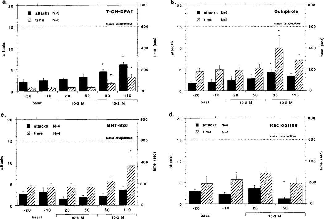
Fig. 1.
Effects of bilateral perfusion with various dopaminergic compounds in the GP/putamen (GP: globus pallidus) on cataplexy in narcoleptic canines. Local perfusion 7-OH-DPAT (10 M) (a), quinpirole (10−4–10−2 M) BHT-920 (10−4–10−2 M) (c) and raclopride M) in the narcoleptic canines is shown. Drugs were mixed into artificial cerebrospinal fluid and perfused through microdialysis probes at the indicated concentrations over the course of a 4-h experiment: none during the first hour,10−4 M during the second hour, 10−3 M during the third hour and 10−2 M during the fourth hour. Each drug was tested with four narcoleptic canines, though the same animals were not always used. The mean number of cataplectic attacks and elapsed time for two food-elicited cataplexy tests (FECT) per test period is shown. For the purpose of figure presentation, status cataplecticus (complete muscle atonia) was arbitrarily designated as 15 attacks elapsed over 600 s. Each drug perfusion time point was compared with the basal time points using a Fisher PLSD post-hoc test; * indicates P < 0.05 satisfactory for comparison with either basal time point. Univariate analyses of number of cataplectic attacks over the tested dose range for each drug were; 7-OH-DPAT:F4,20 = 9.851, P=0.001 Quinpirole: F4,28=3.244, P = 0.048; BHT-920: F4,28 = 1.322, P = 0.279; Raciopride: F2,14=4.235, P = 0.028. Univariate analyses of elapsed FECT time over the tested dose range for each drug were; 7-OH-DPAT: F 4,20= 19.366, P = Quinpirole: F4,28=3.975, P = 0.026; BHT-920: F4,28=4.257, P = 0.006; Raciopride: F2,14=1.112, P = 0.655.

