| Metal ion fluorescent probe |
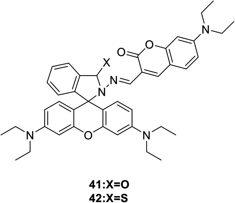
Reacts with Cu2+
|
|
41: Fluorescence changed from yellow to bright red |
3-Position |
| 42: Show strong orange fluorescence at 590 nm |
|
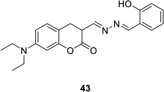
Fluorescence quenching |
3-Position |
|
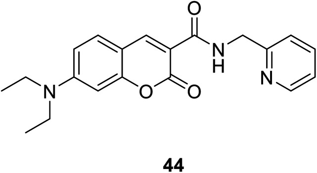
Show fluorescence enhancement |
3-Position |
|
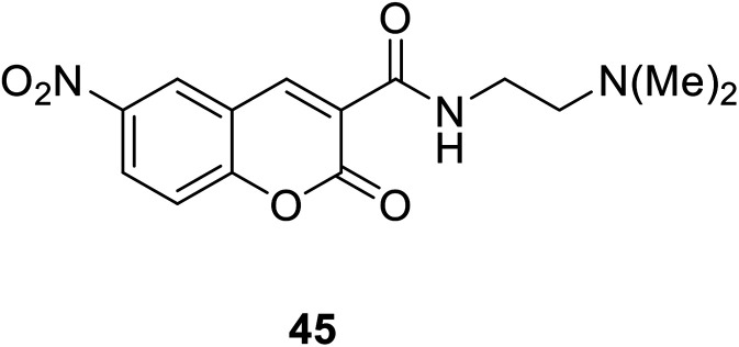
Show fluorescence enhancement |
3-Position |
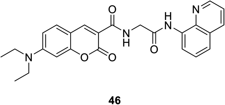
|
“Turn-on” probe; from no fluorescent to fluorescent |
3-Position |
| Reacts with Hg2+
|
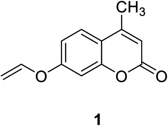
|
Show strong blue fluorescence |
7-Position |
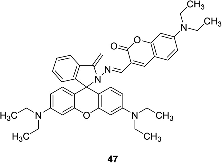
|
Show a reversible dual chromo- and fluorogenic response |
3-Position |
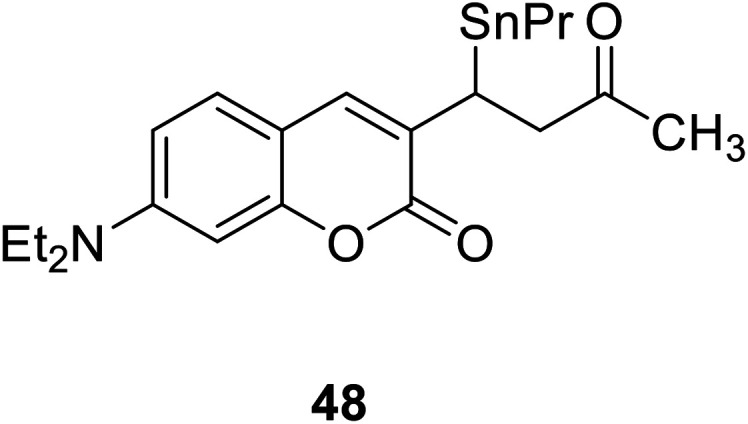
|
Show fluorescence enhancement |
3-Position |
| Reacts with Mg2+
|
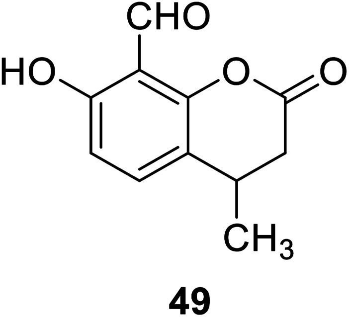
|
Fluorescence changed from weak blue to bright blue |
7-Position and 8-position |
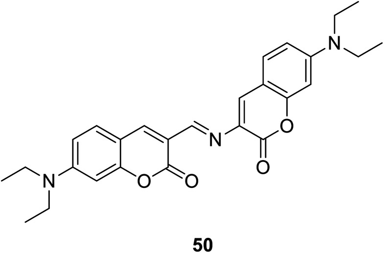
|
Fluorescence changed from non to strong red |
3-Position |
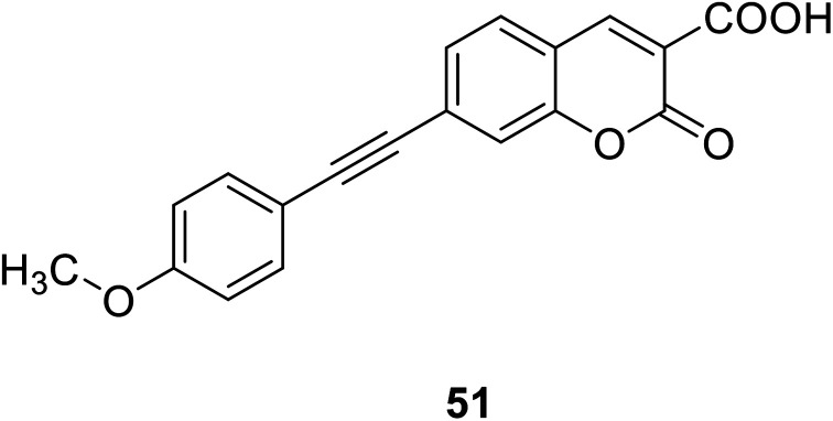
|
Show fluorescence enhancement |
3-Position |
|
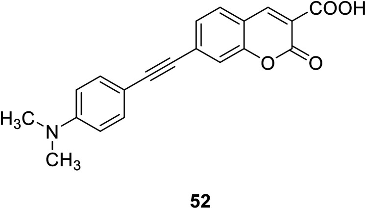
|
Show fluorescence enhancement |
3-Position |
| Reacts with Zn2+
|
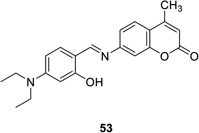
|
Show significant increase in fluorescence at 500 nm |
7-Position |
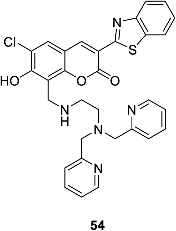
|
Absorption spectrum shifted toward longer wavelengths, and the fluorescence is enhanced |
7-Position and 8-position |
| pH fluorescent probe |
Reacts with H+
|
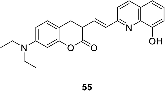
|
As the pH decreased, the absorption spectrum gradually red-shifted |
3-Position |
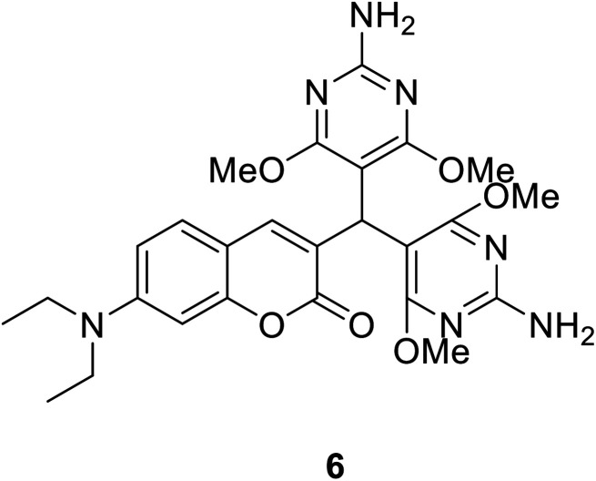
|
When the pH is low (below 3.5), the fluorescence at 460 nm is quenched |
3-Position |
|
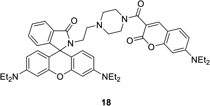
|
When the probe interacted with H+, the coumarin release decreased at 477 nm and the rhodamine release increased at 582 nm |
3-Position |
| Oxygen and sulfide fluorescent probes |
Reacts with oxygen |
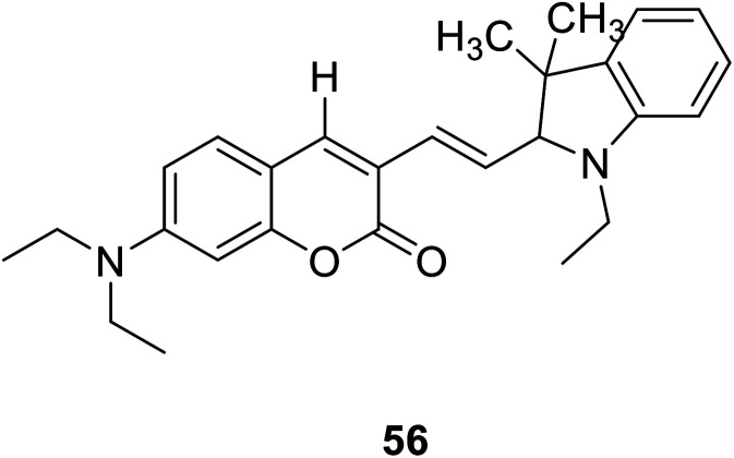
|
I
651/I495 increased significantly |
3-Position |
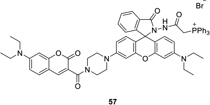
|
New emission appeared at 580 nm. The emission intensity increased with the increase of hypochlorite dose, and the fluorescence intensity decreased at 470 nm |
3-Position |
| Reacts with H2S |
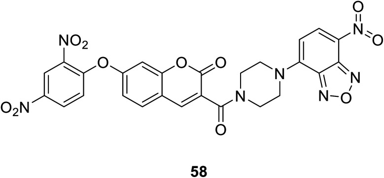
|
Fluorescence quenching |
Functionalized position of fluorescent probe based on FRET mechanism: 3-position |
| Functionalized position of fluorescent probe based on ICT mechanism: 7-position |
|
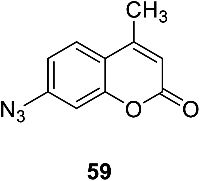
Show fluorescence enhancement |
7-Position |
|
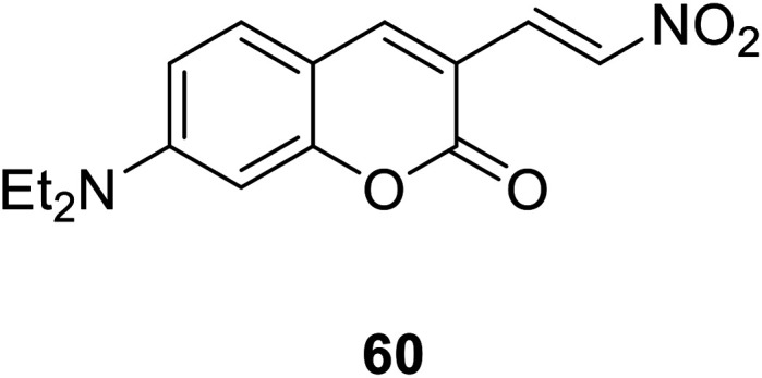
Generates a large emission displacement |
3-Position |
| Environmental polarity probes |
Environmental polarity |
|
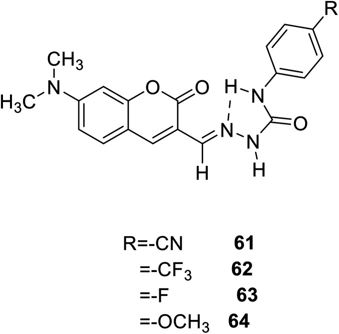
Show orange-yellow colour change and simultaneous fluorescence increase |
3-Position |
|
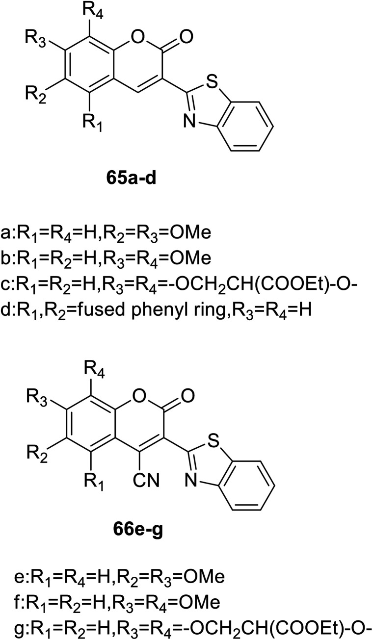
When the polarity of environment decreases, it shows strong fluorescence |
66e–g: 4-position |


