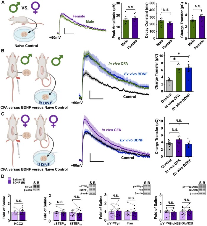
Figure 2.
NMDARs at female SDH synapses are not potentiated or upregulated by the ex vivo BDNF or in vivo CFA models of pathological pain. (A) Baseline lamina I mEPSCs do not differ between male and female adult SD rats. Left: Saline-treated male vs female lamina I neurons, created using BioRender.com. Middle: Average mEPSCs at +60 mV in lamina I neurons from male (green) and female (purple) adult rats. Right: Peak amplitude, decay constant and charge transfer of the NMDAR component of mEPSCs at +60 mV do not differ between male and female SD rats. n = 10 for males and n = 9 for females. Compared using independent samples t-test. (B) Male lamina I NMDAR mEPSCs are potentiated following CFA hindpaw injection and ex vivo BDNF treatment. Left: Experimental paradigm showing male in vivo CFA versus ex vivo BDNF models, created using BioRender.com. Middle: NMDAR mEPSCs from male rat lamina I neurons; control in black, CFA in green, BDNF in blue. Right: Charge transfer of NMDAR mEPSCs for groups shown to left. n = 10 for control, n = 8 for CFA and n = 6 for BDNF. Compared using Welch’s test followed by Games–Howell comparison. (C) Female lamina I NMDAR mEPSCs are not potentiated following CFA hindpaw injection or ex vivo BDNF treatment. Left: Experimental paradigm showing female in vivo CFA versus ex vivo BDNF models, created using BioRender.com. Middle: NMDAR mEPSCs from female rat lamina I neurons; control in black, CFA in purple, BDNF in blue. Right: Charge transfer of NMDAR mEPSCs shown to left. n = 10 for control, n = 8 for CFA and n = 8 for BDNF. Compared using the Kruskal–Wallis one-way ANOVA. (D) Ex vivo BDNF treatment model elicits no effect in female rat SDH synaptosomes. Plots (left) and representative western blots (right) from female rat SDH synaptosomes of tissue treated with either control saline (lilac, n = 8) or 50 ng/ml recombinant BDNF for 70 min (purple, n = 8; compared using paired-samples t-test). *P < 0.05.

