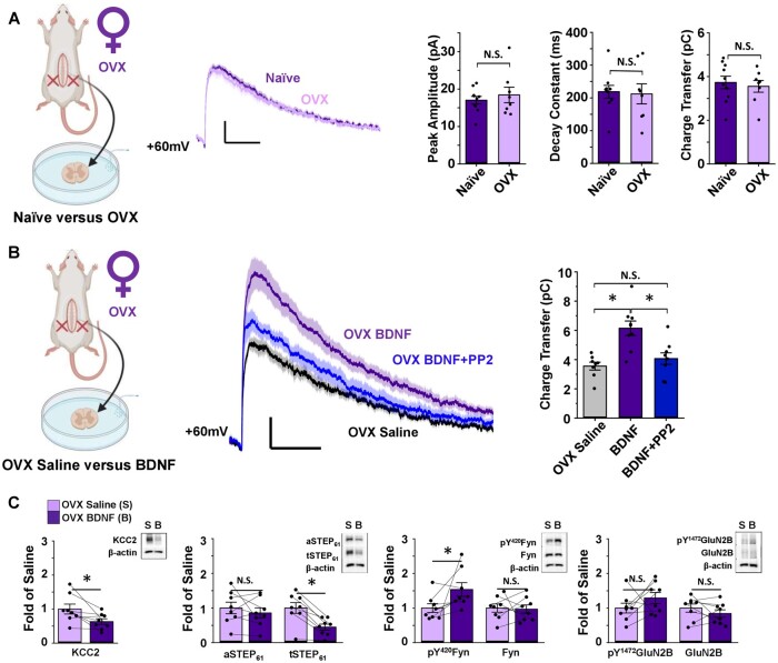
Figure 4.
Ovariectomy triggers BDNF-mediated NMDAR potentiation by the KCC2/STEP61/SFK pathway in SDH neurons of female rats. (A) Ovariectomy (OVX) has no effect on baseline lamina I NMDAR mEPSCs of female SD rats. Left: Saline-treated naïve female versusOVX female lamina I neurons, created using BioRender.com. Middle: Average mEPSCs at +60 mV in lamina I neurons of naïve (purple) and ovariectomized (lilac) female rats. Right: Peak amplitude (compared using Mann–Whitney test), decay constant (compared using independent samples t-test) and charge transfer (compared using independent samples t-test) of the NMDAR component of mEPSCs do not differ between naïve (n = 10) and OVX (n = 8) female rats. (B) In OVX rats, NMDAR mEPSCs in lamina I neurons are potentiated following ex vivo BDNF treatment. This potentiation is blocked using co-treatment with the SFK inhibitor, PP2. Left: Recordings from lamina I neurons were compared for saline- versus BDNF-treated slices from OVX rats, created using BioRender.com. Middle: Average mEPSCs at +60 mV from OVX female rat lamina I neurons; control in black, BDNF in purple, BDNF+PP2 in blue. Right: Charge transfer of NMDAR mEPSCs shown on left. n = 8 for control, n = 9 for BDNF and BDNF + PP2. Comparisons were made using one-way ANOVA, followed by Tukey HSD when P < 0.05. (C) Ex vivo BDNF treatment in OVX rat SDH synaptosomes results in upregulation of pY420Fyn and downregulation of KCC2 and STEP61. Plots (left) and representative western blots (right) from OVX female rat SDH synaptosomes of tissue treated with either control saline (lilac, n = 8) or 50 ng/ml recombinant BDNF for 70 min (purple, n = 8, compared using paired samples t-tests). *P < 0.05.

