Figure 3.
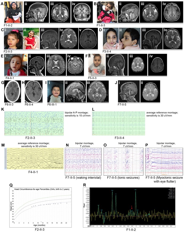
Facial features, brain images, neurophysiologic studies, and head circumference chart of affected individuals from Families 1–7 with biallelic variants in SLC38A3. [A(i)] Photograph of Individual II-2 (Family 1) at 6 years of age. [A(ii–iv)] MRI brain of Individual II-2 from Family 1 at 6 years of age showing foreshortening of corpus callosum (CC), moderate to severe cerebral volume loss, thin brainstem and cervical spine [A(ii) T1 mid-sagittal sequence], under-opercularization and widening Sylvian fissure, abnormal myelination for age [A(iii) T2 axial sequence], mild (infero-lateral), and cerebellar atrophy [A(iv) T1 coronal sequence]. [B(i)] Photograph of Individual II-3 (Family 1) at 4 years of age. [B(ii–iv)] MRI brain of Individual II-3 from Family 1 at 4.5 years of age showing foreshortening of CC, moderate cerebral and mild cerebellar volume loss [B(ii) T1 mid-sagittal sequence], under-opercularization and widening Sylvian fissure, abnormal myelination for age [B(iii) T2 axial sequence, and B(iv); T1 axial sequence]. [C(i and ii)] Photographs of Individual II-3 (Family 2) at 5 years of age. [C(iii–v)] MRI brain of Individual II-3 from Family 2 at 9 months of age showing hypo-intensity of splenium of CC secondary to recent seizure or a remote infarct [C(iii) T1 mid-sagittal sequence], and normal myelination for age [C(iv) T2 axial sequence, and C(v) T1 axial sequence]. [D(i and ii)] Facial photographs of Individual II-4 (Family 3) at 6 years of age. [D(iii and iv)] Brain MRI of Individual II-4 from Family 3 at 2.5 years of age showing mild cerebral volume loss, slight foreshortening of CC [D(iii) T1 mid-sagittal sequence], under-opercularization and widening Sylvian fissure, and delayed myelination for age [D(iv) T2 axial sequence]. [E(i)] Photograph of Individual II-1 (Family 4) at 4 years of age. [E(ii)] Brain MRI (mid-sagittal T1 sequence) of Individual II-1 from Family 4 at 3 years of age showing normal brain structures [E(iii and iv)] axial sequences showing normal myelination for age. [F(i)] Photographs of Individual II-3 (Family 5) at 3 years of age. [F(ii–iv)] MRI brain of Individual II-3 from Family 5 at 21 months of age showing borderline low-normal body of CC, and mild cerebral volume loss [F(ii)] T1 mid-sagittal sequence), under-opercularization and widening Sylvian fissure, and abnormal myelination for age. [F(iii)] T2 axial sequence, and (iv) T1 axial sequence. (G) Head CT of Individual II-3 from Family 6 showing normal brain parenchyma. (H) Head CT of Individual II-4 from Family 6 showing under-opercularization with normal brain parenchyma. [I(i)] Photographs of Individual III-1 (Family 6) at 15 months of age. [I(ii and iii)] MRI brain of Individual III-1 (Family 6) showing mild cerebral atrophy, thin CC, mild superior vermian volume loss and delayed myelination for age [I(ii) T2 mid-sagittal sequence and (iii) T2 axial sequence]. [J(i and ii)] MRI brain of Individual II-5 from Family 7 at 6 months of age showing under-opercularization and widening Sylvian fissure, and abnormal myelination for age [J(i) T1 mid-sagittal sequence, and (ii) T2 axial sequence]. (K) EEG epoch (11 s) of Family 2, Individual II-3 (bipolar montage; sensitivity 15 μV/s) showing background slowing and abundant multifocal epileptiform activity. (L) EEG epoch (17 s) of Family 3, Individual II-4 (average reference montage; sensitivity 30 μV/s) showing background slowing and multifocal epileptiform activity. (M) EEG epoch (10 s) of Family 4, Individual II-1 (average reference montage; sensitivity 30 μV/s) showing a mild background slowing. (N) EEG epoch (10 s) of Family 7, Individual II-5 (bipolar montage; sensitivity 7 μV/s) at 12 months showing diffuse background slowing and near-continuous high amplitude spike and slow-wave epileptiform activity over posterior regions. (O) EEG epoch (11 s) of Family 7, Individual II-5 (bipolar montage; sensitivity 7 μV/s) at 12 months during a tonic seizure. (P) EEG epoch (10 s) of Family 7, Individual II-5 (bipolar montage; sensitivity 7 μV/s) at 12 months during a myoclonic seizure. (Q) CDC head circumference chart from birth to 2 years of age for Family 2, Individual II-3 showing a progressive/postnatal microcephaly pattern. (R) MRS of Family 1, Individual II-2. The red arrow points to the low glutamate-glutamine (Gx) peak. A-P = anterior-posterior.

