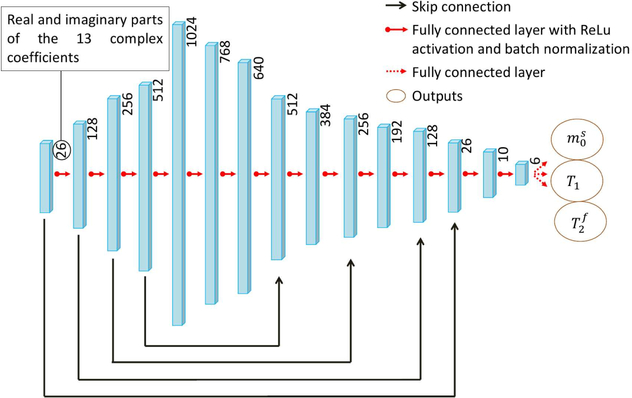
FIGURE 2.
The neural network architecture used in this study. The 13 complex-valued coefficients of each voxel are concatenated and fed into the fully connected network. Skip connections43 are incorporated to avoid the vanishing gradient problem during training. The network outputs the underlying tissue parameters , T1 and , but additional parameters, such as B0 and B1, and be added to the output layer (cf. Supporting Information Figure S4).

