
FIGURE 3. Gut-mesentery-lung axis in Schistosomiasis-associated PAH (Sch-PAH).
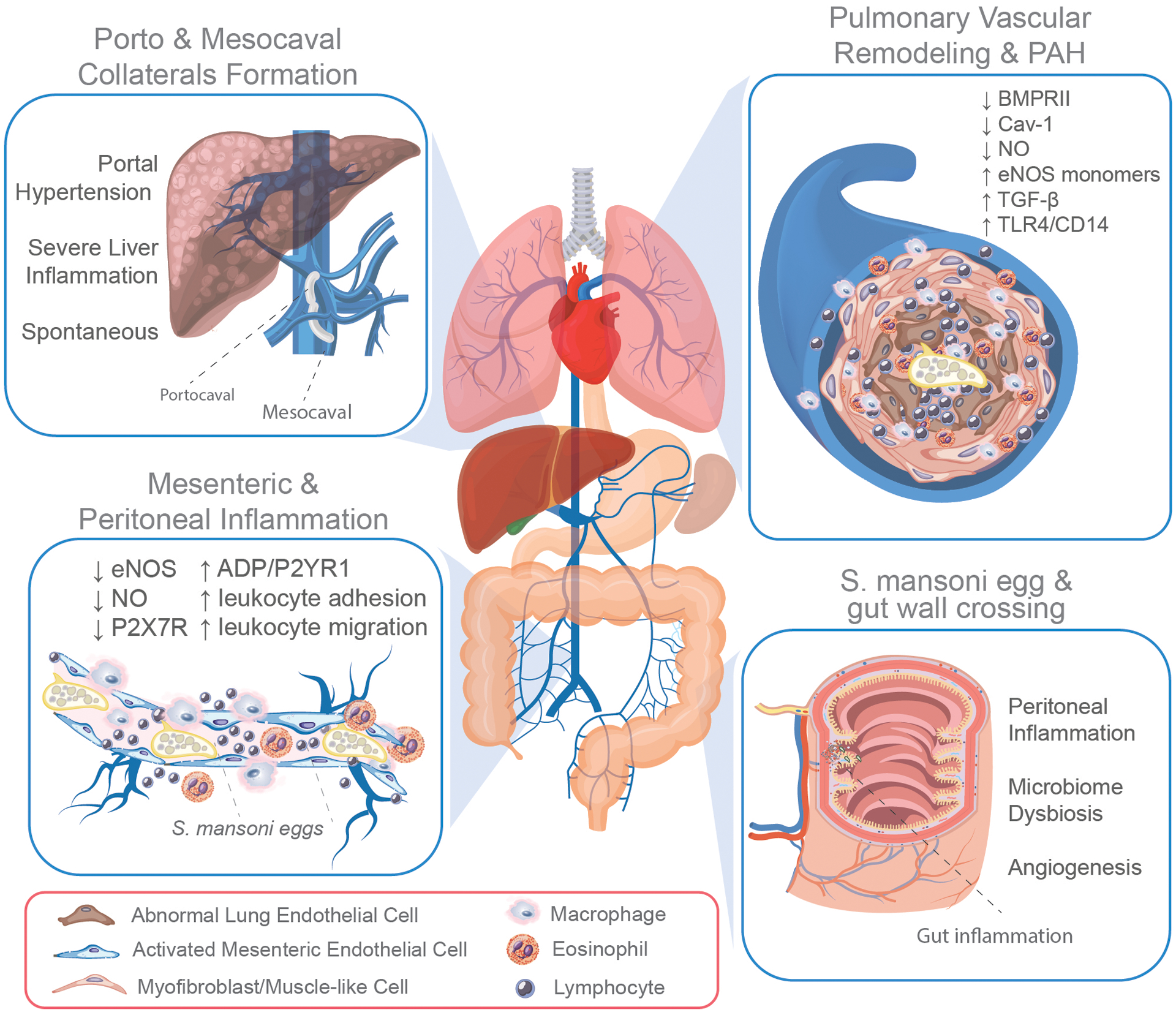
Chronically infected individuals host in their mesenteric system adult schistosomes that lay thousands of eggs. The interaction of adult S. mansoni and eggs with mesenteric endothelial cells contributes to endothelial dysfunction by reducing the expression and function of the endothelial expression of nitric oxide (eNOS) and purinergic receptor P2X7R, and by increasing the ADP-induced activation of P2YR1 and leukocyte adhesion and transmigration into the peritoneal cavity (bottom right box). Once released, half of the eggs actively cross the intestinal wall causing a significant inflammatory response in the gut wall and peritoneal cavity, contributing to microbiome dysbiosis and angiogenesis (bottom left box). The remaining eggs allocate to other organs, including the liver, where they can obstruct the sinusoid vasculature leading to severe inflammation and the development of portal hypertension. In response to elevated portal pressure, the formation of porto and mesocaval collateral circulation not only bypasses the liver but also allows the translocation of S. mansoni eggs, antigens, and toxins into the lungs. Although rare, the formation of spontaneous collateral circulation can also occur (top left box). The translocation of eggs from the mesentery into the lung leads to obliteration, inflammatory remodeling of the pulmonary vasculature, and development of Sch-PAH, which exhibits abnormal endothelial proliferation owing to their transformation into myofibroblasts or mesenchymal-like cells, associated with the reduced expression of bone morphogenetic protein receptor 2 (BMPRII), caveolin-1 (Cav-1), and nitric oxide (NO) and the elevated activation of TGF-β and TLR4/CD14-mediated signaling pathway (top right box).