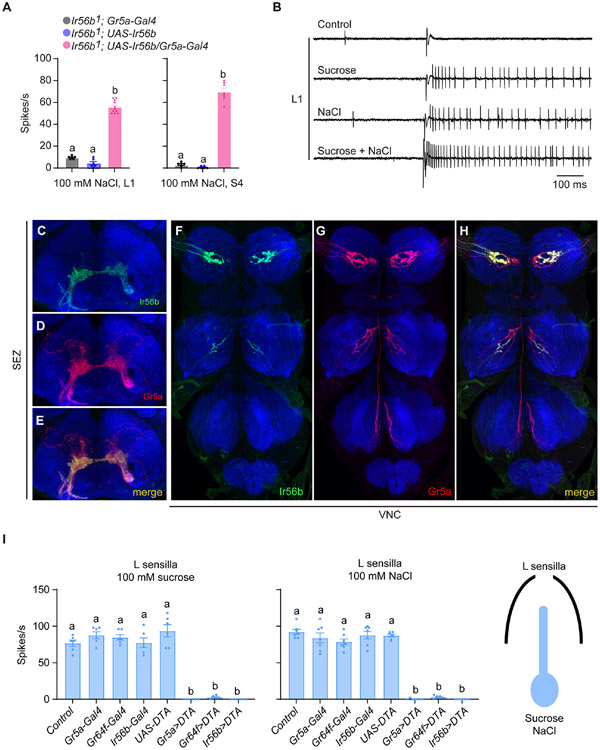
Figure 4. Ir56b is expressed in a subset of sugar-sensitive neurons.
(A) Responses of L1 and S4 in the indicated genotypes to 100 mM NaCl. One-way ANOVA followed by Tukey’s multiple comparison test; n = 6-11. Values indicated with different letters are significantly different.
(B) Sample traces of electrophysiological recordings from L1 in control flies presented with diluent control (30 mM TCC), 50 mM sucrose, 50 mM NaCl, and mixture of 50 mM NaCl and 50 mM sucrose. Sucrose, NaCl, and the mixture were all dissolved in 30 mM TCC.
(C-E) Projection patterns of Ir56a-GAL4- and Gr5a-LexA-expressing neurons in the suboesophageal ganglion (SEZ).
(F-H) Projection patterns of Ir56a-GAL4- and Gr5a-LexA-expressing neurons in the ventral nerve cord (VNC).
(I) Expression of diphtheria toxin under the control of Gr5a-, Gr64f-, or IR56b-Gal4 drivers in L sensilla severely reduced response to both sucrose and NaCl. One-way ANOVA followed by Tukey's multiple comparison test; n=6-7. Values indicated by different letters are different. p<0.05.

