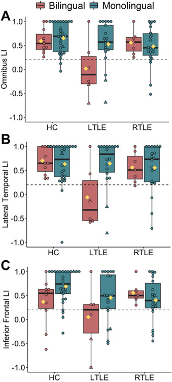
Figure 3. The effect of group on language lateralization is dependent on language status.
fMRI laterality index (LI) plotted separately by group (age-matched controls; HC, left TLE; LTLE, and right TLE; RTLE) and language status (bilingual, monolingual) for A) omnibus language LI, B) inferior frontal LI, and C) lateral temporal LI. Right- and left-handed/ambidextrous individuals are coded as circles and triangles, respectively. Yellow diamonds represent group means. Individuals below the horizontal dotted line have atypical language lateralization (LI ≤ 0.2). Rates of atypical (i.e., right or bilateral) lateralization based on omnibus LI were as follows: monolingual HC=7% (2/29), bilingual HC=0% (0/11), monolingual LTLE=14% (3/21), bilingual LTLE (67%; 4/6), monolingual RTLE=18% (4/22) and bilingual RTLE=0% (0/7). Boxplot denotes the median (bold bar), first and third quartiles (box limits), and ±1.5 times the interquartile range (whiskers).

