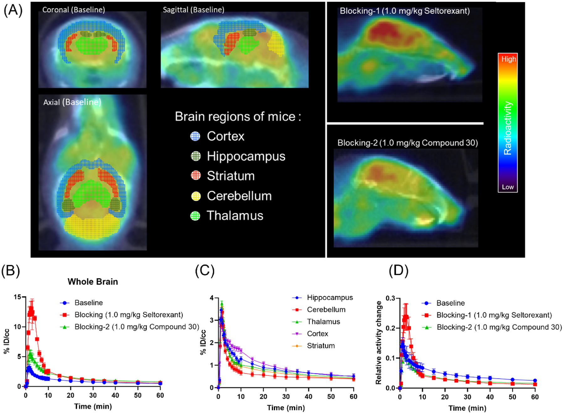
Figure 4.

(A) Representative baseline and blocking PET/CT images of [18F]Seltorexant focus on the mice brain (0–60 min, the baseline is shown as horizontal, coronal, and sagittal plane; the blockings are shown as a sagittal plane); (B) the baseline and blocking time-activity curves of [18F]Seltorexant in the mice whole brain; (C) Time-radioactivity curves for [18F]Seltorexant in the hippocampus, cerebellum, thalamus, cortex, and striatum (n = 3); (D) the normalized baseline and blocking time-activity curves [18F]Seltorexant (normalized the brain uptake curves with the highest radioactivity in the blood at each time point).
