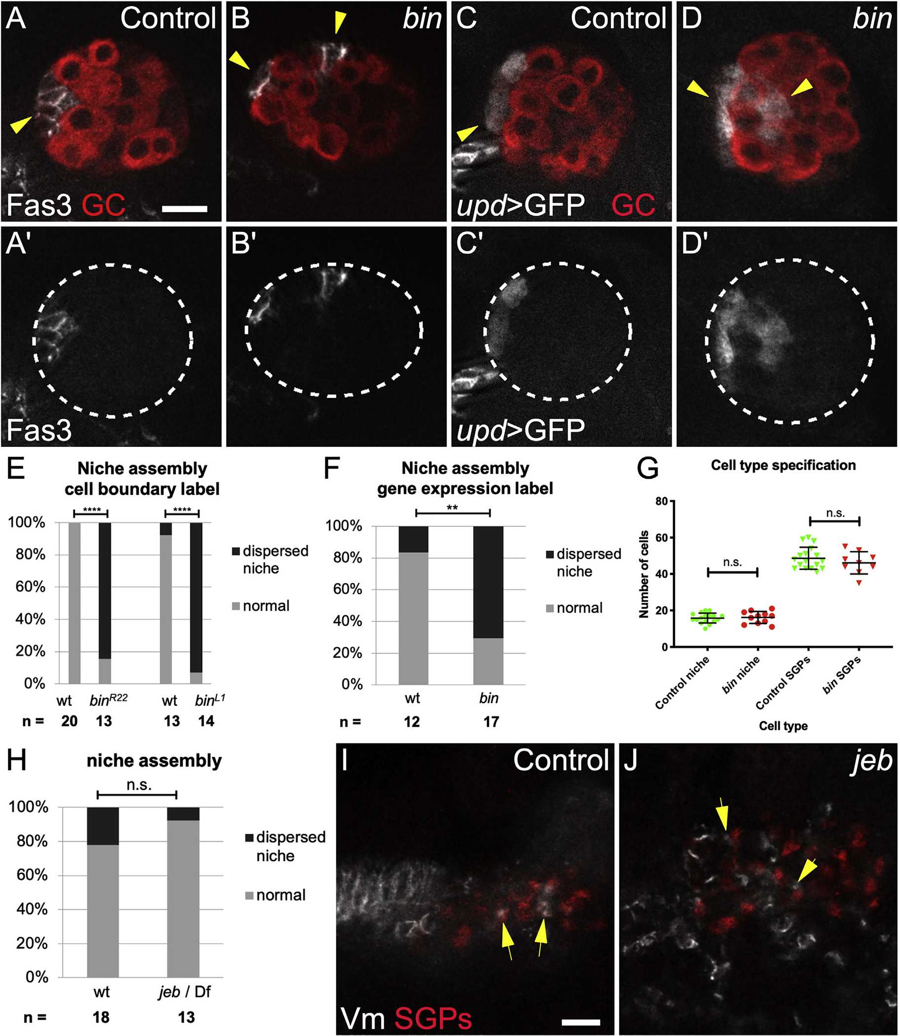
Figure 1. Visceral mesoderm is required for niche assembly and positioning.

(A) Control Stage 17 gonad when niche morphogenesis is complete, immunostained with Vasa (red, germ cells) and Fas3 (white, niche cells, arrow).
(B) biniou mutant; Vasa (red) and Fas3 (white) reveal a dispersed niche (arrowheads).
(C) Control and (D) biniou mutant gonads immunostained with Vasa (red) and expressing upd-Gal4, UAS-GFP in niche cells (white, arrow).
(A′ and B′) Fas3 alone; (C′ and D′) GFP alone. Dotted lines, gonad boundary.
(E and F) Quantification using (E) Fas3 or (F) upd > GFP as marker (p < 0.001, p = 0.004, respectively, Fisher’s exact test).
(G) Number of niche and non-niche SGPs specified in biniou mutants compared with siblings.
(H) Niche assembly is not affected in jeb mutants compared with siblings.
(I) Control and (J) jeb mutant embryos (Stage 13, before gonad coalescence); arrows show SGPs (Traffic jam, red) in contact with Vm cells (Fas3, white). Jeb mutants have a different arrangement of Vm precursors. Scale bars, 10 μm.
