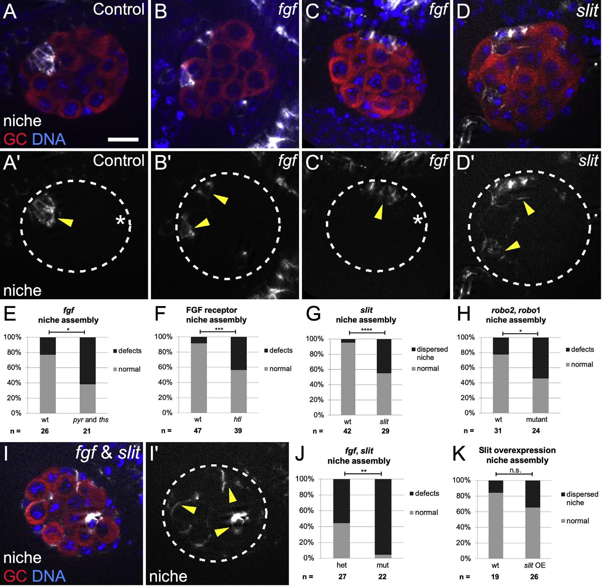
Figure 2. Slit and FGF signaling promote anterior niche assembly.

(A–D and I) Stage 17 gonads, merge of Vasa (red, germ cells), Hoechst (blue, DNA), and Fas3 (white, niche cells), or single channel (Fas3); dotted line, gonad boundary. Scale bars, 10 μm. Prime panels show Fas3 (niche cells, arrowheads) alone.
(A) Sibling controls have a single, anterior niche.
(B and C) Df(2R)BSC25 gonads, with a deletion removing pyr and ths genes, exhibit niche defects such as (B) dispersed niche cell aggregates and (C) niches not at the gonad anterior (asterisk, gonad posterior).
(D) slit[2] mutant gonads often have dispersed niche cell aggregates.
(E–H) Quantification of niche defects (Fisher’s exact test), (E) with pyr and ths removed (fgf) (p = 0.016), (F) FGF htl receptor mutant (p = 0.0003), (G) slit mutant (p < 0.0001), and (H) robo2, robo1 double mutant (p = 0.024).
(I and J) Combined mutant with slit, and pyr and ths removed (fgf) exhibit dispersed niche cells (p = 0.003).
(K) Niche morphogenesis defects were not significant (n.s.) in Slit overexpression embryos.
