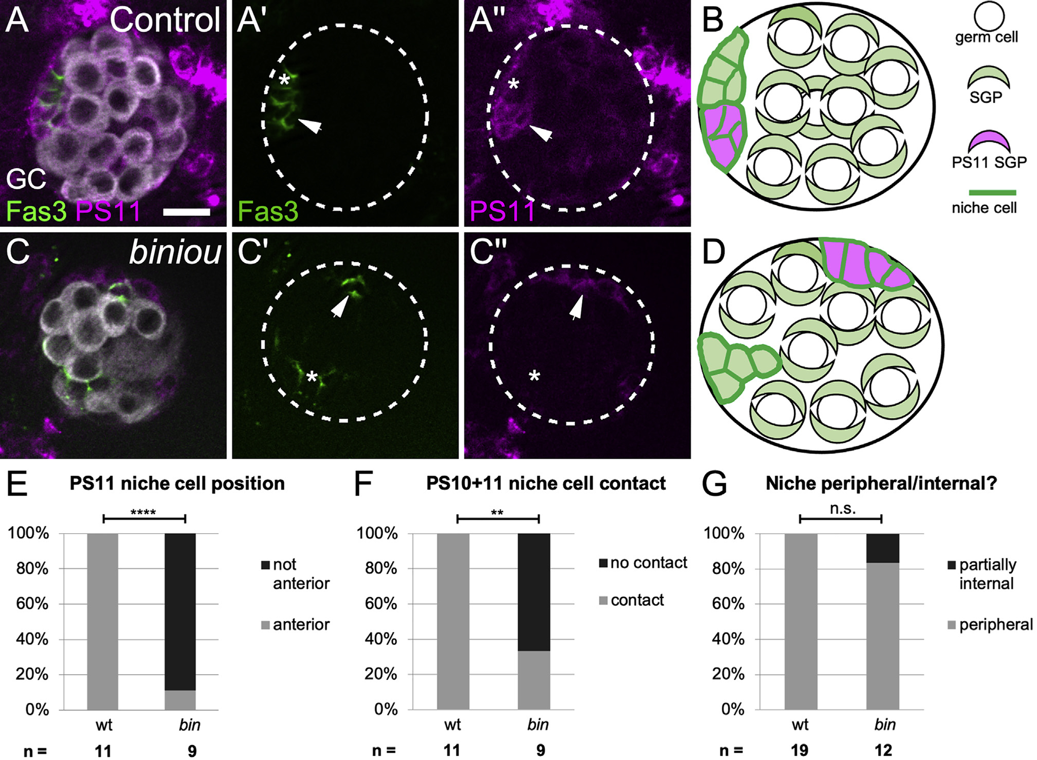
Figure 3. biniou is required for anterior movement of pro-niche cells.

(A and C) Stage 17 gonads expressing mcd8GFP in PS11 cells (magenta), and immunostained with Vasa (white, germ cells) and Fas3 (green, all niche cells). (A) A control, with a single anterior niche (left, green) containing cells deriving from both PS 10 (A′, green alone, asterisk) and PS 11 (A′ and A′′, magenta and green, arrow). (C) biniou mutant with dispersed niche cell aggregates (green). Anterior niche cells deriving from PS 10 (C′, green alone, asterisk) do not associate with PS 11-derived niche cells (C′′, magenta and green, arrows). Ectopic PS11 niche cells were distinguishable from PS13 msSGPs, which do not express the niche cell marker Fas3.
(B and D) Cartoons illustrating the distribution of PS11 niche cells in (B) control and (D) biniou mutants.
(E–G) Quantifications comparing biniou mutants and sibling controls (Fisher’s exact test) by how often (E) PS 11-derived niche cells are located at anterior (p < 0.0001), (F) PS 11 niche cells contact anterior PS 10 niche cells (p < 0.0022), and (G) niche cells are located within 2 μm of the gonad periphery. Scale bars, 10 μm.
