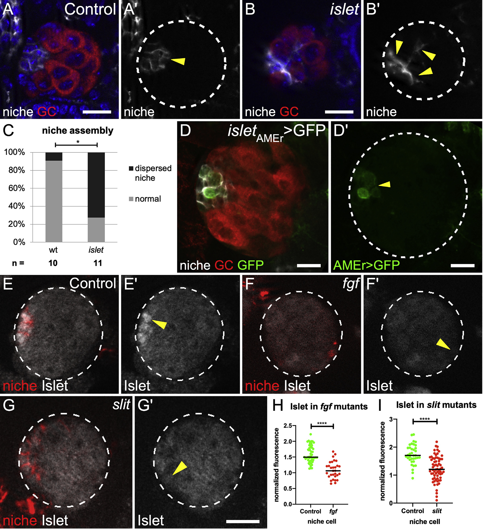
Figure 4. islet is expressed in niche cells in response to Vm signals.

(A and B) (A) Control and (B) islet mutant Stage 17 gonads immunostained with Vasa (red, germ cells), Fas3 (white, niche cells), and Hoechst (blue, nuclei). (A′ and B′) Fas3 alone (arrows).
(C) Quantification of niche assembly in islet versus sibling controls (p = 0.024, Mann-Whitney test).
(D) St 17 gonad expressing GFP driven by the islet AMEr enhancer stained with Vasa (germ cells, red) and Fas3 (niche cells, white). (D′) islet AMEr-driven GFP alone.
(E–G) Stage 17 gonads immunostained for Islet (white), Fas3 (red, niche cells), and Vasa (not shown, germ cells). Gonad boundaries, dotted lines. Arrowheads, niche cells. (E′–G′) Islet alone.
(H and I) Islet accumulation in niche cells from (H) pyr and ths removed (fgf) and (I) slit mutants, compared with sibling controls (p < 0.0001, Mann-Whitney test). Scale bars, 10 μm.
