
Figure 1. EM Identification and 3D reconstruction of neighboring astrocytes.
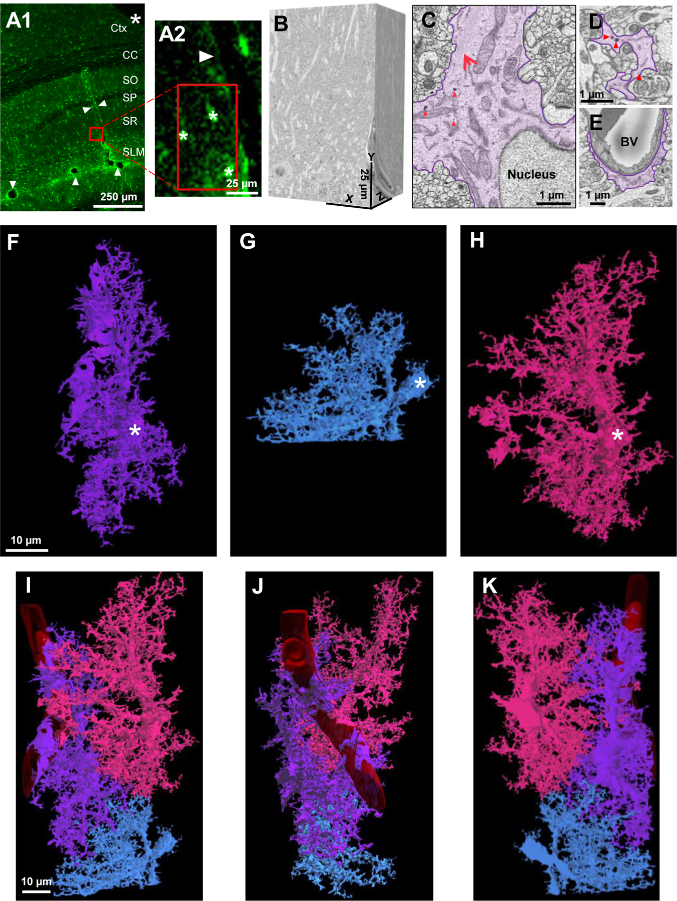
A1) Low magnification confocal microscope image of a fixed brain section displaying the location of eGFP+ astrocytes and delineation of blood vessels (white arrowheads). An angular cut (*) at the upper-right region of the tissue serves as a fiduciary mark. A2) A magnified area from A1 shows the spatial location of 3 neighboring astrocytes next to a blood vessel. Asterisks (*) denote the 3 astrocytes observed in the SBF-SEM images. B) The resulting 500-stack SBF-SEM volume dataset from the selected ROI containing neighboring astrocytes from the stratum radiatum hippocampal subregion. C) An astrocyte was identified by first locating the nucleus-containing cell body. Bundles of filaments (large, red arrow) and several examples of glycogen granules (red arrowheads, also see Fig. S1) are noted near the astrocytic nucleus. D) Astrocyte processes (purple) that extend from the cell body possess an irregular and angular shape. E) Processes that contact blood vessels expand into specialized astrocyte endfeet processes. Abbreviations: Ctx-Cortex, CC-Corpus callosum, SO-Stratum oriens, SP-Stratum pyramidale, SR-stratum radiatum, SLM-stratum lacunosum-moleculare, BV-blood vessel. F-H) 3-dimensional view of three reconstructed astrocytes: purple, blue, and pink. White asterisks found within each cell denote the somas, respectively. I-K) Combined reconstruction depicting the front, side, and back views of the three astrocytes. Each astrocyte is labeled in a different color to clearly demarcate individual astrocyte domains and cellular structures. Note that the blue astrocyte appears ‘smaller’ in size as only part of the cell was included in the EM stack.