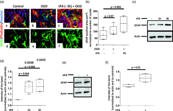
Figure 3.
Preconditioning with tPA induces astrocytic activation. a. The in vitro model of the blood-brain barrier (BBB) was maintained under normoxic conditions (panels a & d), or treated with either PBS (panels b & e) or 5 nM of tPA (panels c & f) three hours before exposure to 60 minutes of OGD conditions. Inserts were then harvested and their underside was stained with phalloidin (red), Hoechst (blue) and anti- GFAP antibodies (green). Confocal micrographs were taken at 60 X magnification. B. GFAP-immunoreactive area normalized to the number of Hoechst-positive nuclei on the underside of the BBB inserts exposed to the experimental conditions described in b. n = 5 inserts per experimental group, assembled with cells from 3 different cultures. Statistical analysis: one way ANOVA with Holm-Sidak’s multiple comparisons. c & d. Representative Western blot analysis (C) and quantification of the intensity of the band normalized to actin (D) of GFAP abundance in astrocytes treated 1 or 3 hours with 5 nM of tPA. n = 4 per experimental group. Statistical analysis: one way ANOVA with Dunnett’s multiple comparisons test. e & f. Representative Western blot analysis (E) and quantification of the intensity of the band normalized to actin (F) of GFAP abundance in astrocytes treated during three hours with vehicle (control) or 5 nM of proteolytically inactive tPA (itPA). n = 4 per experimental group. Statistical analysis: two-tailed student’s t-test.

