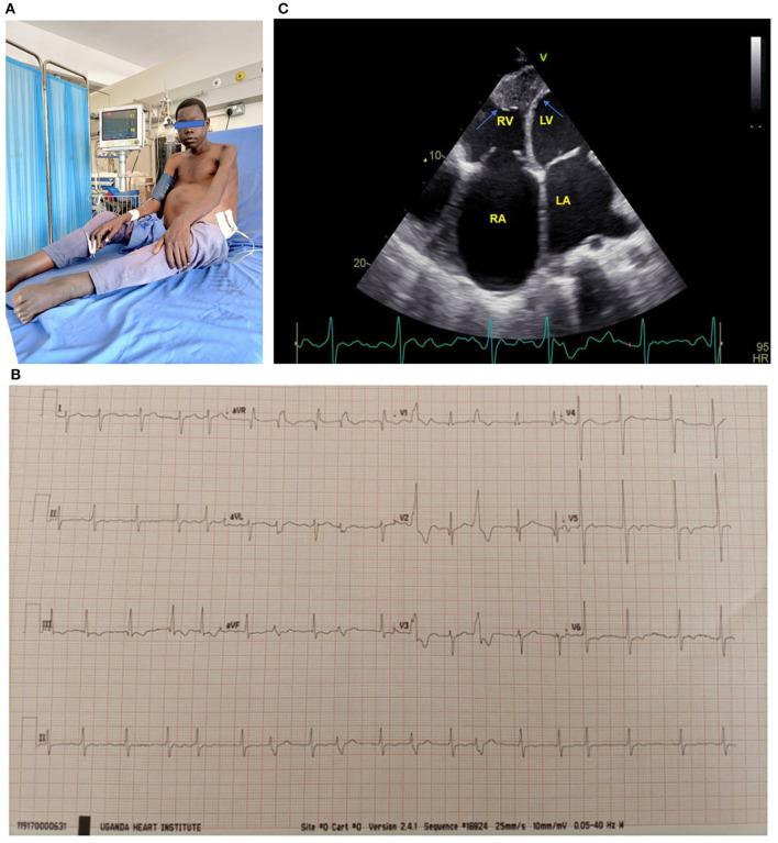
Figure 1.
(A) 17-year-old boy with biventricular EMF receiving paracentesis. Note the gross abdominal ascites. (B) ECG of a 17-year-old boy with biventricular EMF. Note the atrial fibrillation with rapid ventricular response and frequent premature ventricular contractions. (C) Apical 4 chamber view of a 17-year-old boy with biventricular EMF, with predominant right ventricular involvement. Note reduced RV cavity with near obliteration of the apex and fibrotic endocardium (blue arrows) and markedly dilated right atrium. The patient's ECG tracing shows he is in atrial fibrillation. RA, right atrium; RV, right ventricle; LA, left atrium; LV, left ventricle. See the Supplementary Material for more echo images of the above patient.

