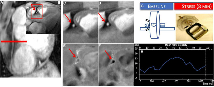
FIGURE 2.
Example of coronary endothelial function (CEF) testing using non-contrast MRI with isometric handgrip exercise (IHE). Scout MRI (A) and cross-sectional cine (B–D) and phase-contrast images (E,F) in a healthy subject showing RCA in cross-section (red arrow). In the expanded inset sections, coronary area increases from rest (C) to IHE (D) and velocity and flow increase from rest (E) to stress (F) (note that increased darkness represents increased signal and thus velocity down through the imaging plane). (G) Stress MRI protocol for CEF measures for endpoints: change in coronary cross sectional area and blood flow velocity (%) from baseline to stress (continuous IHE for 5–8 min). (H) Example coronary flow velocity curve of RCA.

