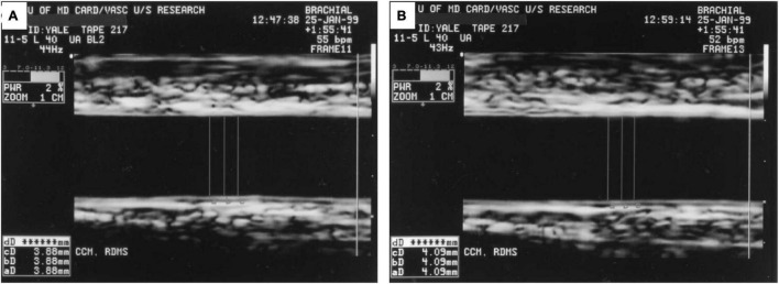
FIGURE 5.
Ultrasound Images Demonstrating Brachial Flow-Mediated Dilatation. (A) Shows the brachial artery at rest with arterial diameter of 3.88 mm. (B) Shows the artery 1 min after hyperemic stimulus with arterial diameter of 4.09 mm. Figure reproduced with permission from Corretti et al. (9) copyright JACC (Elsevier).

