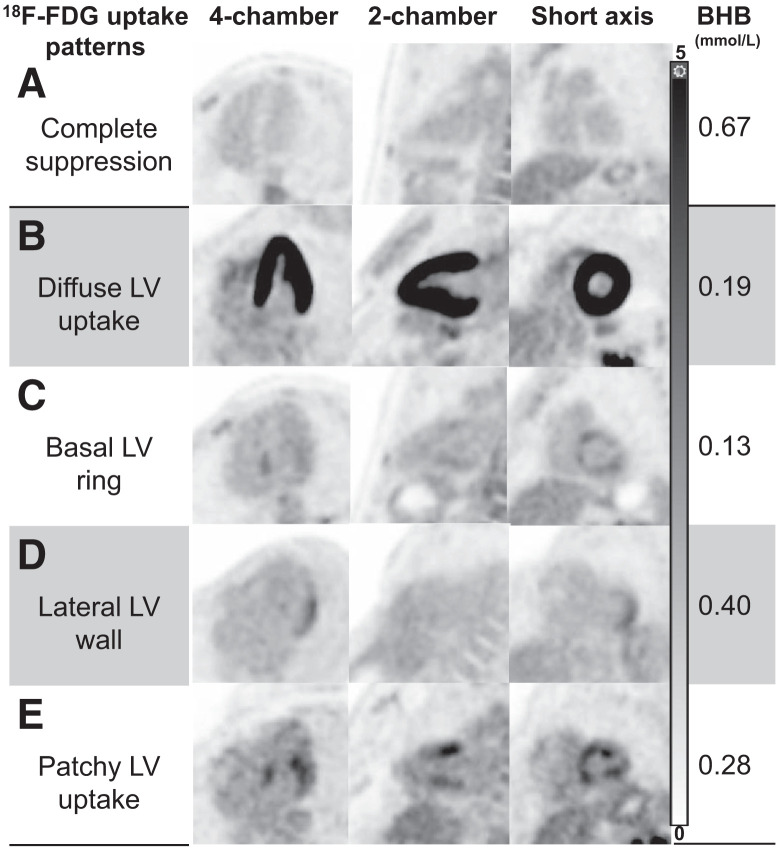
FIGURE 5.
Myocardial 18F-FDG uptake patterns in healthy volunteers. Representative images (displayed using same window width 0–5) of most common myocardial 18F-FDG uptake patterns encountered in our healthy cohort, with their corresponding BHB levels. Patterns C–E can be potentially mistaken as myocardial inflammation; however, accompanying BHB levels should raise concern for incomplete suppression. LV = left ventricular.

