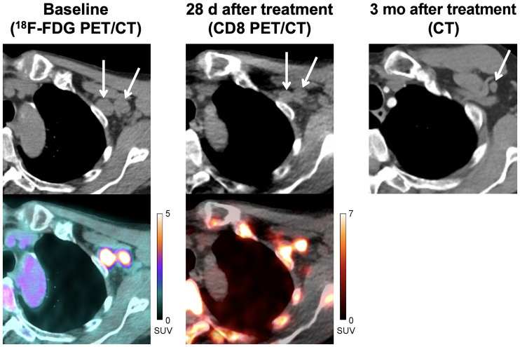
FIGURE 4.
A 71-y-old man with locally advanced stage III melanoma treated with pembrolizumab. Baseline CT and fused 18F-FDG PET/CT images (left) demonstrate 2 18F-FDG–avid metastases in left axilla (SUVMAX = 10.0, medial node; SUVMAX = 7.6, lateral node). CT and fused CD8 PET/CT images (middle) obtained at 28 d after start of immunotherapy demonstrate increased tracer activity in both metastases (SUVMAX = 9.5, medial node; SUVMAX = 10.0, lateral node), suggestive of tumor infiltration by CD8+ T cells. Follow-up imaging with contrast-enhanced CT (right) demonstrated complete response to therapy.

