
FIG. 3.
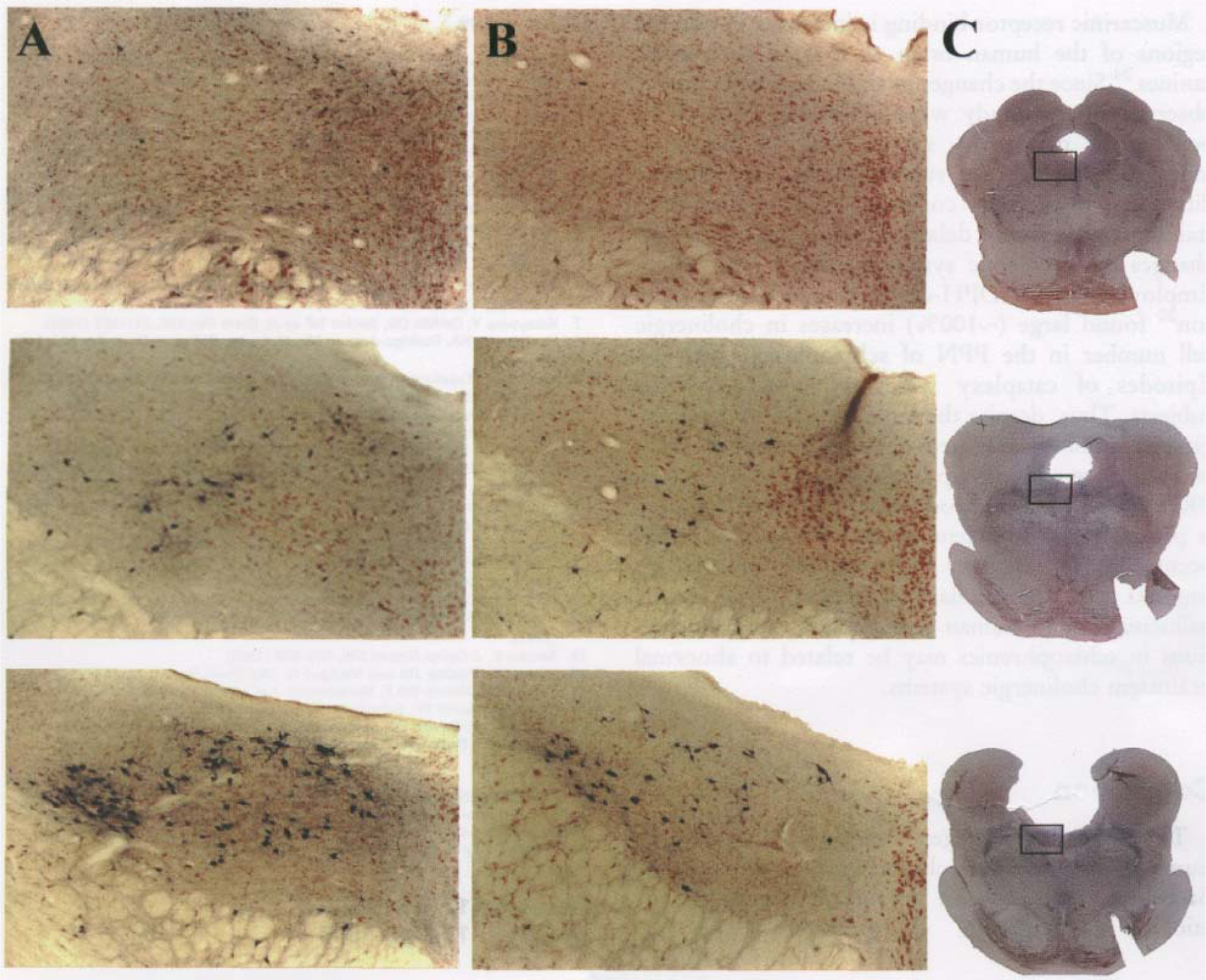
Comparison of NADPH-diaphorase staining in individual LDT nuclei of narcoleptic (A) and normal (B) dogs at brain stem levels (from top to bottom) R9-R8, R8-R7, and R7-R6. Magnification=40x. (C) Low power photographs of normal brain stem slices used in B. The outlined area corresponds to the high power photographs of A and B. Note the greater number of stained cells in the narcoleptic in area R7-R6.